

15 Electron Microscopic in Situ Hybridization and its Combination
213
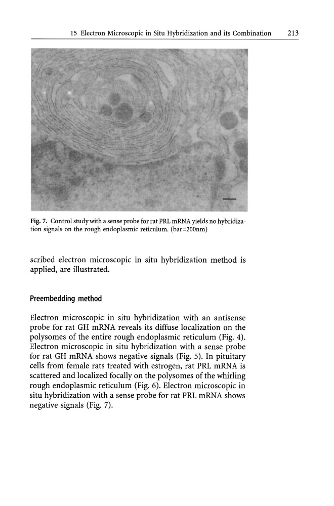
Fig. 7. Control study with a sense probe for rat PRL mRNA yields no hybridization signals on the rough endoplasmic reticulum. (bar=200nm)
scribed electron microscopic in situ hybridization method is
applied, are illustrated.
Preembedding method
Electron microscopic in situ hybridization with an antisense
probe for rat GH mRNA reveals its diffuse localization on the
polysomes of the entire rough endoplasmic reticulum (Fig. 4).
Electron microscopic in situ hybridization with a sense probe
for rat GH mRNA shows negative signals (Fig. 5). In pituitary
cells from female rats treated with estrogen, rat PRL mRNA is
scattered and localized focally on the polysomes of the whirling
rough endoplasmic reticulum (Fig. 6). Electron microscopic in
situ hybridization with a sense probe for rat PRL mRNA shows
negative signals (Fig. 7).
213
Fig. 7. Control study with a sense probe for rat PRL mRNA yields no hybridization signals on the rough endoplasmic reticulum. (bar=200nm)
scribed electron microscopic in situ hybridization method is
applied, are illustrated.
Preembedding method
Electron microscopic in situ hybridization with an antisense
probe for rat GH mRNA reveals its diffuse localization on the
polysomes of the entire rough endoplasmic reticulum (Fig. 4).
Electron microscopic in situ hybridization with a sense probe
for rat GH mRNA shows negative signals (Fig. 5). In pituitary
cells from female rats treated with estrogen, rat PRL mRNA is
scattered and localized focally on the polysomes of the whirling
rough endoplasmic reticulum (Fig. 6). Electron microscopic in
situ hybridization with a sense probe for rat PRL mRNA shows
negative signals (Fig. 7).