

12 In Situ Hybridization for RNA: Nonradioactive Probe: Oligo-DNA Probe
175
tured for 7 mins before hybridization. In some cases, probes
are used as cocktails consisting of equimolar mixtures.
a) Formamide is deionized with mixed bed resin.
- The blocking solution containing 500 Ilg/ml normal sheep
IgG, 5% BSA, 0.3 M NaCI, 100 Ilg/ml tRNA, 100 Ilg/ml salmon
testis DNA in PBS
- The chromogen solution containing 3,3' -diaminobenzidine
(DAB), HzOz, nickel and cobalt; 0.5 mg/ml DAB/O.025%
CoClz/0.02% NiS04 (NH4hS04/0.01% HzOz/O.l M phosphate
buffer (pH 7.5)
Procedure
Unless otherwise specified, all steps are performed at room temperature.
The tissues are fixed in 4% PF A/PBS, and embedded in paraffin
according to a routine procedure. The specimens containing
bone or teeth have to be decalcified before being processed to
usual histochemical techniques. The influence of decalcification
procedure on the retention ofhybridizable RNA is an important
consideration. Therefore, to achieve an optimal balance between
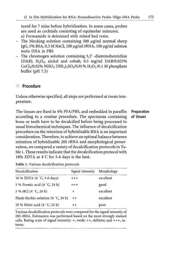
retention of hybridizable 28S rRNA and morphological preservation, we compared a variety of decalcification protocols in Table 1. These results indicate that the decalcification protocol with
10% EDTA at 4°C for 5-6 days is the best.
Table 1. Various decalcification protocols
Decalcification
Signal intensity Morphology
10 % EDTA (4 ce, 5-6 days)
+++
excellent
5 % Formic acid (4 °e, 24 h)
+++
good
5 % Hel (4 °e, 24 h)
+
excellent
Plank-Rychlo solution (4 °e, 24 h) ++
excellent
10 % Nitric acid (4 °e, 24 h)
++
poor
Various decalcification protocols were compared for the signal intenstiy of
28S rRNA. Estimation was performed based on the most strongly stained
cells. Rating scale of signal intensity: +, weak; ++, definite; and +++, intense.
Preparation
of tissues
175
tured for 7 mins before hybridization. In some cases, probes
are used as cocktails consisting of equimolar mixtures.
a) Formamide is deionized with mixed bed resin.
- The blocking solution containing 500 Ilg/ml normal sheep
IgG, 5% BSA, 0.3 M NaCI, 100 Ilg/ml tRNA, 100 Ilg/ml salmon
testis DNA in PBS
- The chromogen solution containing 3,3' -diaminobenzidine
(DAB), HzOz, nickel and cobalt; 0.5 mg/ml DAB/O.025%
CoClz/0.02% NiS04 (NH4hS04/0.01% HzOz/O.l M phosphate
buffer (pH 7.5)
Procedure
Unless otherwise specified, all steps are performed at room temperature.
The tissues are fixed in 4% PF A/PBS, and embedded in paraffin
according to a routine procedure. The specimens containing
bone or teeth have to be decalcified before being processed to
usual histochemical techniques. The influence of decalcification
procedure on the retention ofhybridizable RNA is an important
consideration. Therefore, to achieve an optimal balance between
retention of hybridizable 28S rRNA and morphological preservation, we compared a variety of decalcification protocols in Table 1. These results indicate that the decalcification protocol with
10% EDTA at 4°C for 5-6 days is the best.
Table 1. Various decalcification protocols
Decalcification
Signal intensity Morphology
10 % EDTA (4 ce, 5-6 days)
+++
excellent
5 % Formic acid (4 °e, 24 h)
+++
good
5 % Hel (4 °e, 24 h)
+
excellent
Plank-Rychlo solution (4 °e, 24 h) ++
excellent
10 % Nitric acid (4 °e, 24 h)
++
poor
Various decalcification protocols were compared for the signal intenstiy of
28S rRNA. Estimation was performed based on the most strongly stained
cells. Rating scale of signal intensity: +, weak; ++, definite; and +++, intense.
Preparation
of tissues