

1 Introduction to the Detection of DNA Strand Breaks
9
ferase (TdT). When a tissue section is reacted with TdT in the
presence of biotin-16-dUTP or digoxigenin-ll-dUTP, the sites
of DNA strand breaks can be labeled with these haptenic nucleotide analogues. Finally, the haptens are detected immunohistochemically with enzyme-labeled antibodies such as HRP-Iabeled
anti-biotin.
In tissue sections, differing from the case in solution, the sites
of DSB are preferentially reacted with the enzyme compared to
that ofSSB. Thus, TUNEL is now the most frequently used method to detect apoptotic cells which have abundant DSB of DNA.
Here, however, I would like to raise a warning that TUNEL does
not always detect apoptotic cells. As Bortner et al. (1995) reviewed, it is not prerequisite for apoptotic cells to show a significant level of DNA double-stranded breaks at an internucleosomal site. It is also significant that TdT in itself is not specific to the
3' -OH terminus of DSB, rather it can even react efficiently with
,. .
. . -
• 1 ~
o
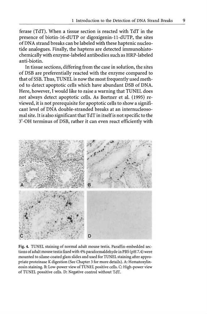
Fig. 4. TUNEL staining of normal adult mouse testis. Paraffin-embedded sections of adult mouse testis fixed with 4% paraformaldehyde in PBS (pH 7.4) were
mounted to silane-coated glass slides and used for TUNEL staining after appropriate proteinase K digestion (See Chapter 3 for more details). A: Hematoxylineosin staining. B: Low-power view ofTUNEL positive cells. C: High-power view
of TUNEL possitive cells. D: Negative control without TdT.
9
ferase (TdT). When a tissue section is reacted with TdT in the
presence of biotin-16-dUTP or digoxigenin-ll-dUTP, the sites
of DNA strand breaks can be labeled with these haptenic nucleotide analogues. Finally, the haptens are detected immunohistochemically with enzyme-labeled antibodies such as HRP-Iabeled
anti-biotin.
In tissue sections, differing from the case in solution, the sites
of DSB are preferentially reacted with the enzyme compared to
that ofSSB. Thus, TUNEL is now the most frequently used method to detect apoptotic cells which have abundant DSB of DNA.
Here, however, I would like to raise a warning that TUNEL does
not always detect apoptotic cells. As Bortner et al. (1995) reviewed, it is not prerequisite for apoptotic cells to show a significant level of DNA double-stranded breaks at an internucleosomal site. It is also significant that TdT in itself is not specific to the
3' -OH terminus of DSB, rather it can even react efficiently with
,. .
. . -
• 1 ~
o
Fig. 4. TUNEL staining of normal adult mouse testis. Paraffin-embedded sections of adult mouse testis fixed with 4% paraformaldehyde in PBS (pH 7.4) were
mounted to silane-coated glass slides and used for TUNEL staining after appropriate proteinase K digestion (See Chapter 3 for more details). A: Hematoxylineosin staining. B: Low-power view ofTUNEL positive cells. C: High-power view
of TUNEL possitive cells. D: Negative control without TdT.