

152
E. MULLER
cell [13, 14]. In order to obtain the actual substrate concentration, it must
not be overlooked that the concentration of urea inside the cell is lower
than in the ambient solution in which small slices of tissue are suspended.
This is a consequence of urea metabolism in the cells, especially at low
overall urea concentrations [14, 15].
The determination of the specific activity or of the turnover number of
an enzyme requires a knowledge of the amount of enzyme per mg protein
(cf. [5]), quantities which cannot be obtained in vivo. Therefore, it remains
only to measure the enzyme activity after extraction under standardized
conditions in vitro [9]. But how can we be sure that the extraction of the
enzyme is quantitatively complete, especially in experiments with plants?
In tackling this problem we focussed our interest onto the Michaelis
constant in vivo, since here the specific activity need not be known; it is
sufficient to compare the reaction rate over a wide range of substrate
concentrations with the reaction rate at saturation. The great advantage is
that the Michaelis constant of urease is identical with the dissociation constant of the enzyme-sub strate-complex and not connected with the overall
reaction rate [11] ; furthermore the constant is independent of the PH ([11] and
[19], but cf. [3] and [17]); consequently, it is not necessary to know the PH
In VIVO.
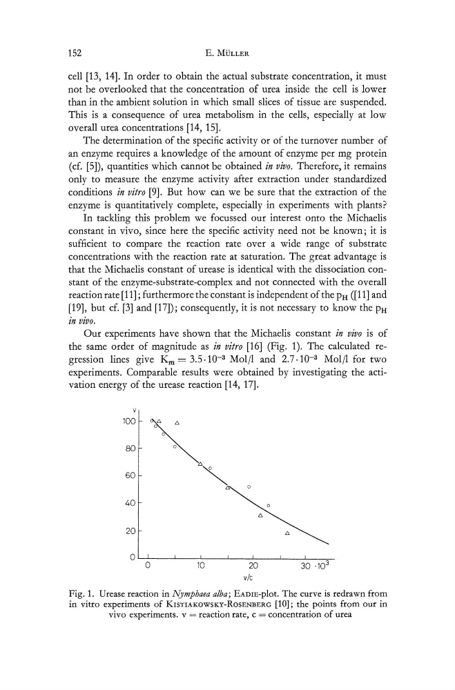
Our experiments have shown that the Michaelis constant in vivo is of
the same order of magnitude as in vitro [16] (Fig. 1). The calculated regression lines give Km = 3.5.10- 3 Molll and 2.7.10- 3 Molll for two
experiments. Comparable results were obtained by investigating the activation energy of the urease reaction [14, 17].
100
~
80
' "
60
~ 0
40
to.
20
to.
0 0
10
20
30 .10 3
vic
Fig. 1. Urease reaction in Nymphaea alba; EADIE-plot. The curve is redrawn from
in vitro experiments of KISTIAKOWSKy-RoSENBERG [10]; the points from our in
vivo experiments. v = reaction rate, c = concentration of urea
E. MULLER
cell [13, 14]. In order to obtain the actual substrate concentration, it must
not be overlooked that the concentration of urea inside the cell is lower
than in the ambient solution in which small slices of tissue are suspended.
This is a consequence of urea metabolism in the cells, especially at low
overall urea concentrations [14, 15].
The determination of the specific activity or of the turnover number of
an enzyme requires a knowledge of the amount of enzyme per mg protein
(cf. [5]), quantities which cannot be obtained in vivo. Therefore, it remains
only to measure the enzyme activity after extraction under standardized
conditions in vitro [9]. But how can we be sure that the extraction of the
enzyme is quantitatively complete, especially in experiments with plants?
In tackling this problem we focussed our interest onto the Michaelis
constant in vivo, since here the specific activity need not be known; it is
sufficient to compare the reaction rate over a wide range of substrate
concentrations with the reaction rate at saturation. The great advantage is
that the Michaelis constant of urease is identical with the dissociation constant of the enzyme-sub strate-complex and not connected with the overall
reaction rate [11] ; furthermore the constant is independent of the PH ([11] and
[19], but cf. [3] and [17]); consequently, it is not necessary to know the PH
In VIVO.
Our experiments have shown that the Michaelis constant in vivo is of
the same order of magnitude as in vitro [16] (Fig. 1). The calculated regression lines give Km = 3.5.10- 3 Molll and 2.7.10- 3 Molll for two
experiments. Comparable results were obtained by investigating the activation energy of the urease reaction [14, 17].
100
~
80
' "
60
~ 0
40
to.
20
to.
0 0
10
20
30 .10 3
vic
Fig. 1. Urease reaction in Nymphaea alba; EADIE-plot. The curve is redrawn from
in vitro experiments of KISTIAKOWSKy-RoSENBERG [10]; the points from our in
vivo experiments. v = reaction rate, c = concentration of urea