

368
ROBERT F. REILLY
Procedure
The reticulocyte lysate system
Reticulocyte lysate is prepared from rabbits made anemic by repeated injection of phenyl-hydrazine (3). Blood is collected, washed, and lysed in
water. After a high speed spin the supernatant is collected. Endogenaus globin mRNA is removed by abrief treatment with calcium-dependent micrococcal nuclease, and the nuclease is subsequently inactivated by chelation of
divalent cations with EDTA (3). This procedure does not alter the protein
synthetic activity of the extract. The requirement for the removal of endogenaus mRNA is the major disadvantage ofthis system. If significant levels
of mRNA remain after the digestion, including sequences that contain initiation sites, exogenaus mRNAs will have to compete with these fragments
for ribosomal binding sites. Weakly initiating mRNA species may do so inefficiently. Unfortunately, the activity of commercially available translation
systems is variable and is never specified for a given lot. The lysate is optimized by the addition of an energy generating system (phosphocreatine
kinase and phosphocreatine), a mixture of tRNAs to expand the range
of mRNAs that can be translated, and hemin to prevent inhibition of initiation (4, 5) (hemin is a suppressor of an inhibitor of the initiation factor
eiF2a). The lysate should be stored in liquid nitrogen since it slowly loses
activity even at -70°C. It should also be stored in aliquots, since it dramatically loses activity on repetitive freeze-thawing.
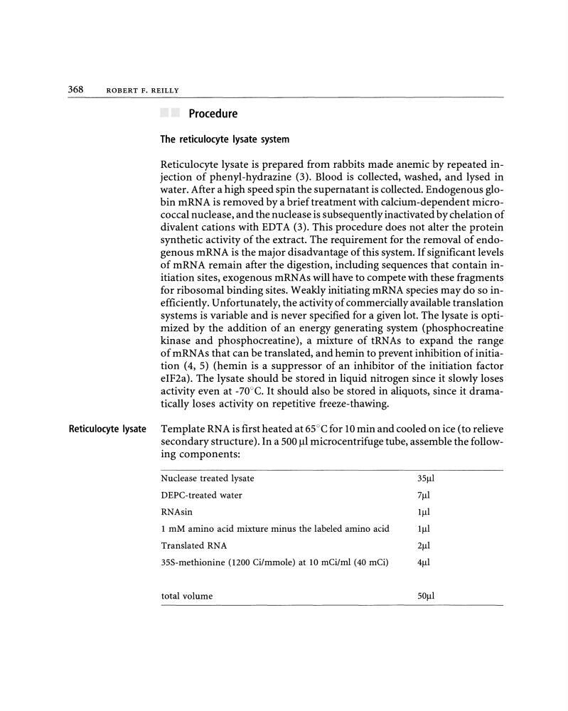
Reticulocyte lysate Template RNA is first heated at 65°C for 10 min and cooled on ice (to relieve
secondary structure). In a 500 Jll microcentrifuge tube, assemble the following components:
Nuclease treated lysate
35J.1l
DEPC-treated water
7J.1l
RNAsin
11-11
1 mM amino acid mixtureminus the labeled amino acid
11-11
Translated RNA
21-11
35S-methionine (1200 Ci/mmole) at 10 mCi/ml (40 mCi)
4Jll
total volume
SOJ.1l
ROBERT F. REILLY
Procedure
The reticulocyte lysate system
Reticulocyte lysate is prepared from rabbits made anemic by repeated injection of phenyl-hydrazine (3). Blood is collected, washed, and lysed in
water. After a high speed spin the supernatant is collected. Endogenaus globin mRNA is removed by abrief treatment with calcium-dependent micrococcal nuclease, and the nuclease is subsequently inactivated by chelation of
divalent cations with EDTA (3). This procedure does not alter the protein
synthetic activity of the extract. The requirement for the removal of endogenaus mRNA is the major disadvantage ofthis system. If significant levels
of mRNA remain after the digestion, including sequences that contain initiation sites, exogenaus mRNAs will have to compete with these fragments
for ribosomal binding sites. Weakly initiating mRNA species may do so inefficiently. Unfortunately, the activity of commercially available translation
systems is variable and is never specified for a given lot. The lysate is optimized by the addition of an energy generating system (phosphocreatine
kinase and phosphocreatine), a mixture of tRNAs to expand the range
of mRNAs that can be translated, and hemin to prevent inhibition of initiation (4, 5) (hemin is a suppressor of an inhibitor of the initiation factor
eiF2a). The lysate should be stored in liquid nitrogen since it slowly loses
activity even at -70°C. It should also be stored in aliquots, since it dramatically loses activity on repetitive freeze-thawing.
Reticulocyte lysate Template RNA is first heated at 65°C for 10 min and cooled on ice (to relieve
secondary structure). In a 500 Jll microcentrifuge tube, assemble the following components:
Nuclease treated lysate
35J.1l
DEPC-treated water
7J.1l
RNAsin
11-11
1 mM amino acid mixtureminus the labeled amino acid
11-11
Translated RNA
21-11
35S-methionine (1200 Ci/mmole) at 10 mCi/ml (40 mCi)
4Jll
total volume
SOJ.1l