

324
ECKART SCHOTT, MARTIN PAUL AND DETLEV GANTEN
examination is carried out with a speculum; mice are examined using small
forceps. Signs that copulation has taken place are:
• wide vaginal introitus
• introitus plastered by secretions
• copulatory plug in the vagina, which is of white color and adheres tightly
to the wall.
Collection of oocytes
After the animals are killed, the oviduct is removed surgically, the swollen
ampulla is opened, oocytes are collected and immediately placed into tissue-culture medium (M2 medium, (13)) containing Hyaluronidase (4000
IU/ ml) to loosen the adhering cumulus cells. Oocytes are incubated in
this medium for 30 seconds to 3 minutes. After the conglomerate has dissolved, the oocytes are washed 2 to 3 tim es in M2 medium and transferred to
pre-warmed M 16 medium ( 13) in a humidified tissue culture incubator with
So/o C02 at 37oC.
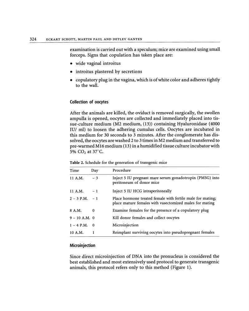
Table 2. Schedule for the generation of transgenic mice
Time
Day
11 A.M.
-3
11 A.M.
- 1
2- 3 P.M. - 1
8 A.M.
0
9- 10 A.M. 0
1-4 P.M. 0
10A.M.
Microinjection
Procerlure
Inject 5 IU pregnant mare serum gonadotropin (PMSG) into
peritoneum of donor mice
Inject 5 IU HCG intraperitoneally
Place hormone treated female with fertile male for mating;
place mature females with vasectomized males for mating
Examine females for the presence of a copulatory plug
Kill donor females and collect oocytes
Microinjection
Reimplant surviving oocytes into pseudopregnant females
Since direct microinjection of DNA into the pronucleus is considered the
best established and most extensively used protocol to generate transgenic
animals, this protocol refers only to this method (Figure 1).
ECKART SCHOTT, MARTIN PAUL AND DETLEV GANTEN
examination is carried out with a speculum; mice are examined using small
forceps. Signs that copulation has taken place are:
• wide vaginal introitus
• introitus plastered by secretions
• copulatory plug in the vagina, which is of white color and adheres tightly
to the wall.
Collection of oocytes
After the animals are killed, the oviduct is removed surgically, the swollen
ampulla is opened, oocytes are collected and immediately placed into tissue-culture medium (M2 medium, (13)) containing Hyaluronidase (4000
IU/ ml) to loosen the adhering cumulus cells. Oocytes are incubated in
this medium for 30 seconds to 3 minutes. After the conglomerate has dissolved, the oocytes are washed 2 to 3 tim es in M2 medium and transferred to
pre-warmed M 16 medium ( 13) in a humidified tissue culture incubator with
So/o C02 at 37oC.
Table 2. Schedule for the generation of transgenic mice
Time
Day
11 A.M.
-3
11 A.M.
- 1
2- 3 P.M. - 1
8 A.M.
0
9- 10 A.M. 0
1-4 P.M. 0
10A.M.
Microinjection
Procerlure
Inject 5 IU pregnant mare serum gonadotropin (PMSG) into
peritoneum of donor mice
Inject 5 IU HCG intraperitoneally
Place hormone treated female with fertile male for mating;
place mature females with vasectomized males for mating
Examine females for the presence of a copulatory plug
Kill donor females and collect oocytes
Microinjection
Reimplant surviving oocytes into pseudopregnant females
Since direct microinjection of DNA into the pronucleus is considered the
best established and most extensively used protocol to generate transgenic
animals, this protocol refers only to this method (Figure 1).