

12 Chromosome Analysis by Fluorescence in situ Hybridization
167
The combined use of several probes modified with different reporter systems in a single experiment allows the simultaneous visualization of multiple nucleic acid targets within the same preparation and significantly increases the utility of FISH.
Many fields of biomedical research have been greatly influenced by applications of FISH including cytogenetics, gene mapping and cell biology.
The implications of these applications are reviewed in detail elsewhere (Joos
1994; Bentz et al.1994; LeBeau 1993). In this chapter, we describe a basic
protocol for FISH to chromosomal DNA.
Components of a FISH experiment
• Specimens: For most applications, conventional methanol/acetic acid
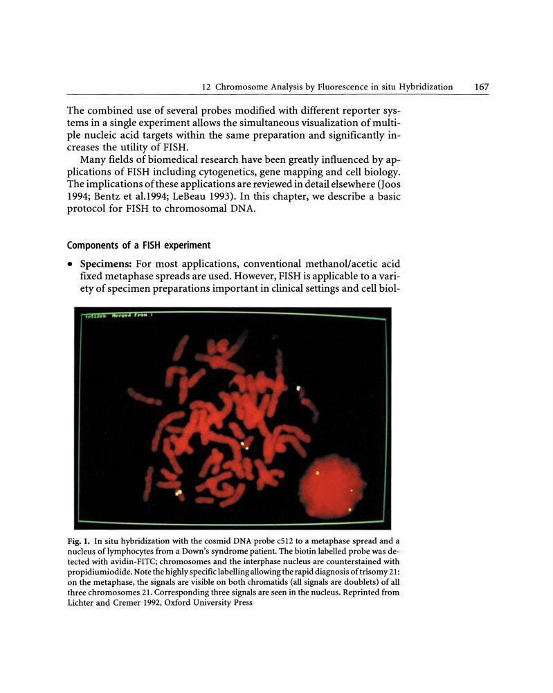
fixed metaphase spreads are used. However, FISH is applicable to a variety of specimen preparations important in clinical settings and cell biolFig. 1. In situ hybridization with the cosmid DNA probe c512 to a metaphase spread and a
nudeus oflymphocytes from a Down's syndrome patient. The biotin Iabelied probe was detected with avidin-FITC; chromosomes and the interphase nudeus are counterstained with
propidiumiodide. Note the highly specific labelling allowing the rapid diagnosis of trisomy 21:
on the metaphase, the signals arevisible on both chromatids (all signals are doublets) of all
three chromosomes 21. Corresponding three signals are seen in the nudeus. Reprinted from
Lichterand Cremer 1992, Oxford University Press
167
The combined use of several probes modified with different reporter systems in a single experiment allows the simultaneous visualization of multiple nucleic acid targets within the same preparation and significantly increases the utility of FISH.
Many fields of biomedical research have been greatly influenced by applications of FISH including cytogenetics, gene mapping and cell biology.
The implications of these applications are reviewed in detail elsewhere (Joos
1994; Bentz et al.1994; LeBeau 1993). In this chapter, we describe a basic
protocol for FISH to chromosomal DNA.
Components of a FISH experiment
• Specimens: For most applications, conventional methanol/acetic acid
fixed metaphase spreads are used. However, FISH is applicable to a variety of specimen preparations important in clinical settings and cell biolFig. 1. In situ hybridization with the cosmid DNA probe c512 to a metaphase spread and a
nudeus oflymphocytes from a Down's syndrome patient. The biotin Iabelied probe was detected with avidin-FITC; chromosomes and the interphase nudeus are counterstained with
propidiumiodide. Note the highly specific labelling allowing the rapid diagnosis of trisomy 21:
on the metaphase, the signals arevisible on both chromatids (all signals are doublets) of all
three chromosomes 21. Corresponding three signals are seen in the nudeus. Reprinted from
Lichterand Cremer 1992, Oxford University Press