

7 Introduction to Pulsed-Field Gel Electrophoresis (PFGE)
Probe A
Probes 8 and C
A
R
p
c N N/R N/C R p c N N/R N/C
= = = = = = = = = = = =
- -
-
- - -
- - - - -
-
-
-
8
Marker A
Marker B
Marker C
c
PIR Nl
•
I I
L..l.
N
1 11~
(C)
R
j
R
-
..
Nru I
- -
(R)
- -
Not I
(N)
Cla I
(C)
Pvu I
(P)
Not/Nru
- -
-
Not/Cia
- -
(C)=incomplete digestion
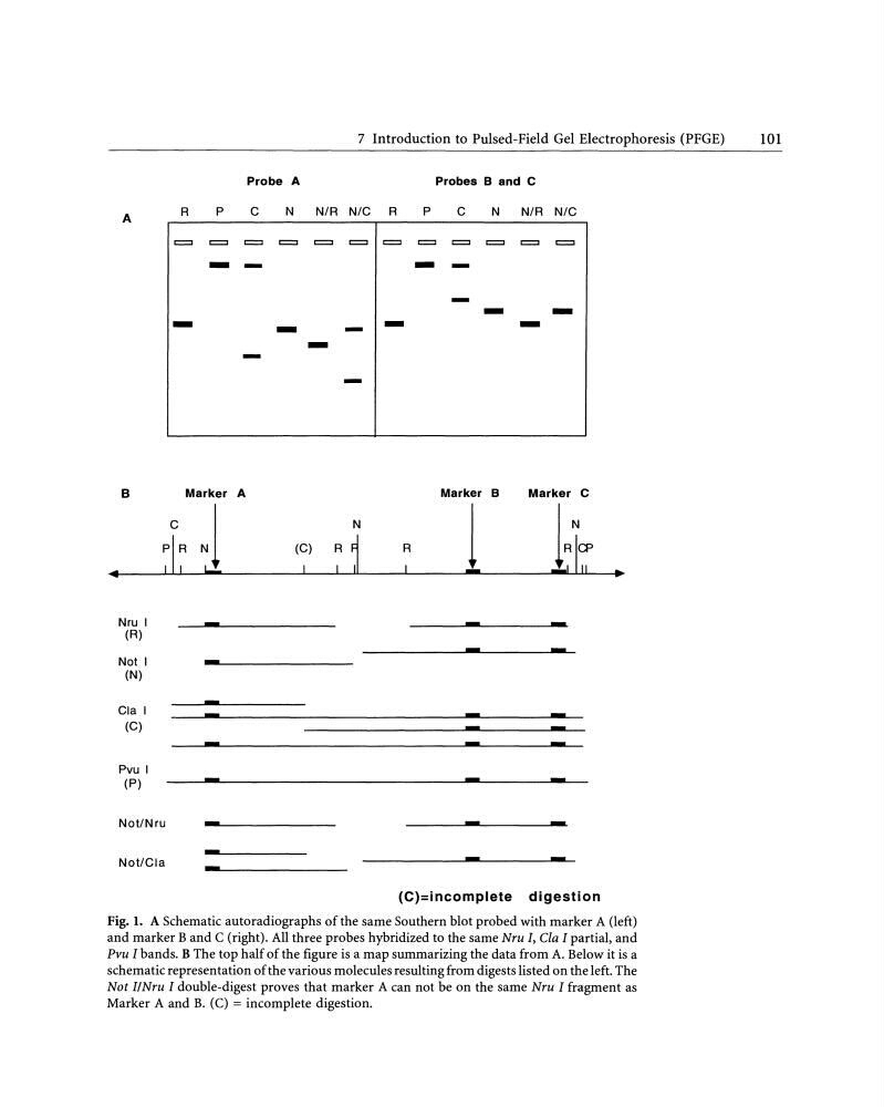
Fig. 1. A Schematic autoradiographs of the same Southern blot probed with marker A (left)
and marker B and C (right). All three probes hybridized to the same Nru I, Cla I partial, and
Pvu I bands. B The top half of the figure is a map summarizing the data from A. Below it is a
schematic representation of the various molecules resulting from digests listed on the left. The
Not 1/Nru I double-digest proves that marker A can not be on the same Nru I fragment as
Marker A and B. (C) = incomplete digestion.
101
Probe A
Probes 8 and C
A
R
p
c N N/R N/C R p c N N/R N/C
= = = = = = = = = = = =
- -
-
- - -
- - - - -
-
-
-
8
Marker A
Marker B
Marker C
c
PIR Nl
•
I I
L..l.
N
1 11~
(C)
R
j
R
-
..
Nru I
- -
(R)
- -
Not I
(N)
Cla I
(C)
Pvu I
(P)
Not/Nru
- -
-
Not/Cia
- -
(C)=incomplete digestion
Fig. 1. A Schematic autoradiographs of the same Southern blot probed with marker A (left)
and marker B and C (right). All three probes hybridized to the same Nru I, Cla I partial, and
Pvu I bands. B The top half of the figure is a map summarizing the data from A. Below it is a
schematic representation of the various molecules resulting from digests listed on the left. The
Not 1/Nru I double-digest proves that marker A can not be on the same Nru I fragment as
Marker A and B. (C) = incomplete digestion.
101