

106
D.M. Paterson and S.E. Hagerthey
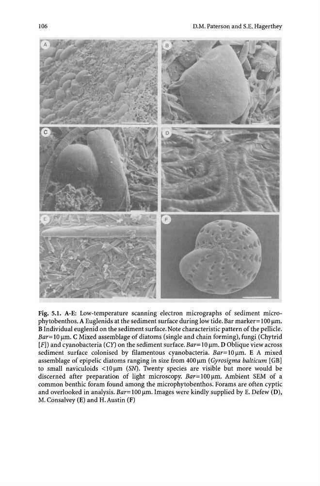
Fig. 5.1. A-E: Low-temperature scanning electron micrographs of sediment microphytobenthos. A Euglenids at the sediment surface during low tide. Bar marker = 100 flm.
B Individual euglenid on the sediment surface. Note characteristic pattern of the pellicle.
Bar= 10 flm. C Mixed assemblage of diatoms (single and chain forming), fungi (Chytrid
[F]) and cyanobacteria (Cy) on the sediment surface.Bar=10 flm. D Oblique view across
sediment surface colonised by filamentous cyanobacteria. Bar= 10 flm. E A mixed
assemblage of epipelic diatoms ranging in size from 400 flm (Gyrosigma balticum [GB]
to small naviculoids <10 flm (SN). Twenty species are visible but more would be
discerned after preparation of light microscopy. Bar= 100 flm. Ambient SEM of a
common benthic foram found among the microphytobenthos. Forams are often cyptic
and overlooked in analysis. Bar= 100 flm. Images were kindly supplied by E. Defew (D),
M. Consalvey (E) and H.Austin (F)
D.M. Paterson and S.E. Hagerthey
Fig. 5.1. A-E: Low-temperature scanning electron micrographs of sediment microphytobenthos. A Euglenids at the sediment surface during low tide. Bar marker = 100 flm.
B Individual euglenid on the sediment surface. Note characteristic pattern of the pellicle.
Bar= 10 flm. C Mixed assemblage of diatoms (single and chain forming), fungi (Chytrid
[F]) and cyanobacteria (Cy) on the sediment surface.Bar=10 flm. D Oblique view across
sediment surface colonised by filamentous cyanobacteria. Bar= 10 flm. E A mixed
assemblage of epipelic diatoms ranging in size from 400 flm (Gyrosigma balticum [GB]
to small naviculoids <10 flm (SN). Twenty species are visible but more would be
discerned after preparation of light microscopy. Bar= 100 flm. Ambient SEM of a
common benthic foram found among the microphytobenthos. Forams are often cyptic
and overlooked in analysis. Bar= 100 flm. Images were kindly supplied by E. Defew (D),
M. Consalvey (E) and H.Austin (F)