

146
Exercise 10
Whipple grid, or plankton counting square), is installed in the plane of the real image
in the ocular (Fig. 10.4). As a result, the image of the specimen being examined appears
to be immediately under the markings on the ocular micrometer. Using the
micrometer, the dimensions of objects can be measured directly.
The Whipple ocular micrometer contains an accurately ruled grid that is subdivided
into 100 squares (Fig. 10.4). One of the central squares is subdivided further into 25
smaller squares. The ocular micrometer is installed by carefully unscrewing the upper
lens mounting of the ocular (usually the right one of a binocular microscope) and
then placing the reticule on the circular diaphragm found about halfway down in
the ocular. After the ocular is reasembled, observe the markings; when they are not
in focus, remove the reticule and reverse it. Using a stage micrometer (Fig. 10.5),
determine the dimensions of the ocular micrometer grid according to the procedure
Whipple Square as -
/~III~
seen Uvough ocu lar
~
("Whipple field")
"Sma II squares" sub tend
~ 01 larlle squares:
5 . 2~
.. Larlle square" subtends
one tenth of entire Whipple
Square: 26~
1-_------1[-- Apparent lines of siehl
subtend 260~ on staae
micrometer scale
\""
't-- lOO~ ----\
_ - - - PORTION OF MAGNIFIED IMAGE OF STAGE MICROMETER SCALE - - - - -
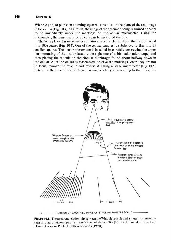
Figure 10.6. The apparent relationship between the Whipple reticule and a stage micrometer as
seen through a microscope at a magnification of about 430 x (lOx ocular and 43 x objective).
[From American Public Health Association (1989).J
Exercise 10
Whipple grid, or plankton counting square), is installed in the plane of the real image
in the ocular (Fig. 10.4). As a result, the image of the specimen being examined appears
to be immediately under the markings on the ocular micrometer. Using the
micrometer, the dimensions of objects can be measured directly.
The Whipple ocular micrometer contains an accurately ruled grid that is subdivided
into 100 squares (Fig. 10.4). One of the central squares is subdivided further into 25
smaller squares. The ocular micrometer is installed by carefully unscrewing the upper
lens mounting of the ocular (usually the right one of a binocular microscope) and
then placing the reticule on the circular diaphragm found about halfway down in
the ocular. After the ocular is reasembled, observe the markings; when they are not
in focus, remove the reticule and reverse it. Using a stage micrometer (Fig. 10.5),
determine the dimensions of the ocular micrometer grid according to the procedure
Whipple Square as -
/~III~
seen Uvough ocu lar
~
("Whipple field")
"Sma II squares" sub tend
~ 01 larlle squares:
5 . 2~
.. Larlle square" subtends
one tenth of entire Whipple
Square: 26~
1-_------1[-- Apparent lines of siehl
subtend 260~ on staae
micrometer scale
\""
't-- lOO~ ----\
_ - - - PORTION OF MAGNIFIED IMAGE OF STAGE MICROMETER SCALE - - - - -
Figure 10.6. The apparent relationship between the Whipple reticule and a stage micrometer as
seen through a microscope at a magnification of about 430 x (lOx ocular and 43 x objective).
[From American Public Health Association (1989).J