

144
Exercise 10
can be viewed with a conventional microscope. The simple apparatus consists of a
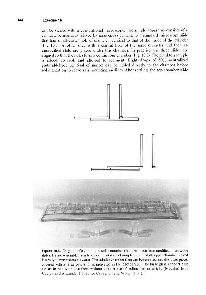
cylinder, permanently affixed by glass epoxy cement, to a standard microscope slide
that has an ofT-center hole of diameter identical to that of the inside of the cylinder
(Fig. 10.3). Another slide with a central hole of the same diameter and then an
unmodified slide are placed under this chamber. In practice, the three slides are
aligned so that the holes form a continuous chamber (Fig. 10.3). The plankton sample
is added, covered, and allowed to sediment. Eight drops of 50% neutralized
glutaraldehyde per 5 ml of sample can be added directly to the chamber before
sedimentation to serve as a mounting medium. After settling, the top chamber slide
Figure 10.3. Diagram of a compound sedimentation chamber made from modified microscope
slides. Upper: Assembled, ready for sedimentation of sample. Lower: With upper chamber moved
laterally to remove excess water. The tubular chamber then can be removed and the lower pieces
covered with a large coverslip, as indicated in the photograph. The large glass support base
assists in removing chambers without disturbance of sedimented materials. [Modified from
Coulon and Alexander (1972): see Crumpton and Wetzel (1981).J
Exercise 10
can be viewed with a conventional microscope. The simple apparatus consists of a
cylinder, permanently affixed by glass epoxy cement, to a standard microscope slide
that has an ofT-center hole of diameter identical to that of the inside of the cylinder
(Fig. 10.3). Another slide with a central hole of the same diameter and then an
unmodified slide are placed under this chamber. In practice, the three slides are
aligned so that the holes form a continuous chamber (Fig. 10.3). The plankton sample
is added, covered, and allowed to sediment. Eight drops of 50% neutralized
glutaraldehyde per 5 ml of sample can be added directly to the chamber before
sedimentation to serve as a mounting medium. After settling, the top chamber slide
Figure 10.3. Diagram of a compound sedimentation chamber made from modified microscope
slides. Upper: Assembled, ready for sedimentation of sample. Lower: With upper chamber moved
laterally to remove excess water. The tubular chamber then can be removed and the lower pieces
covered with a large coverslip, as indicated in the photograph. The large glass support base
assists in removing chambers without disturbance of sedimented materials. [Modified from
Coulon and Alexander (1972): see Crumpton and Wetzel (1981).J