

596
Frederichs et al.
1000
2D
600
E
400
SD
.s
(J)
200
~ 100
Cl
c
0
60
...J
40
20
SP
10
GeoB 1713-5
2- 3cm
<>
<>
SD
<>
2D
<>
SP
GeoB 1713-5
5-6cm
<>
1000
600
400
200
100
60
40
20
10
0.0
0.2
0.4
0.6
0.8
1.00.0
0.2
0.4
0.6
0.8
1.0
Axial Ratio
Axial Ratio
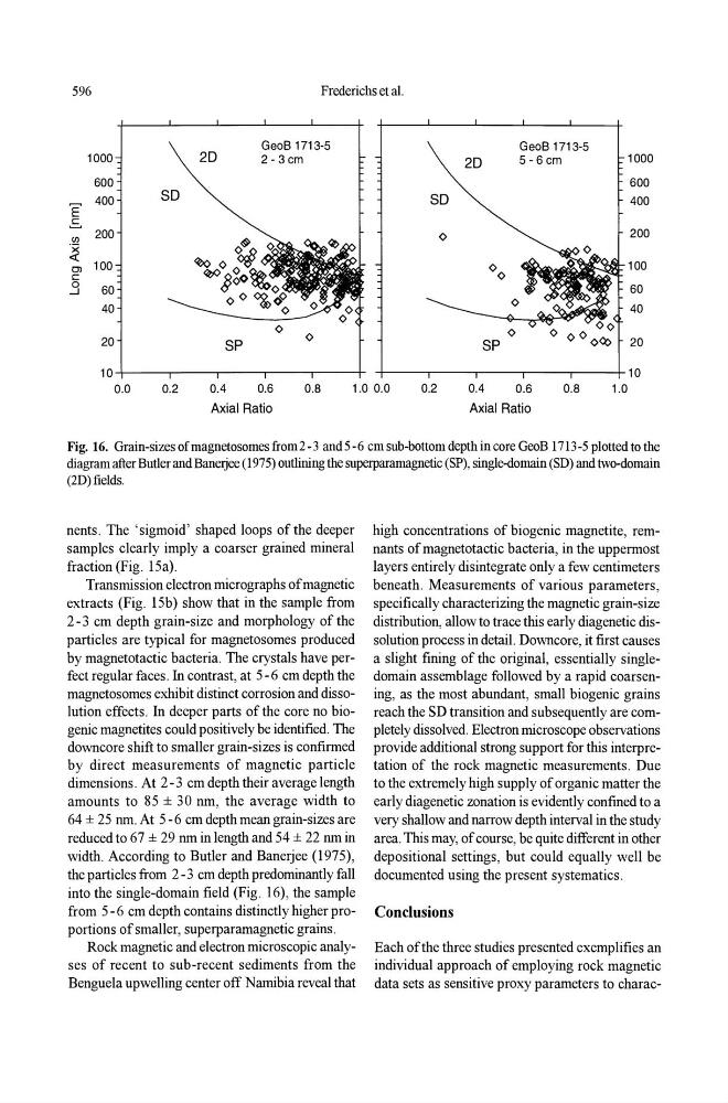
Fig. 16. Grain-sizes of magneto somes from 2 -3 and 5 -6 cm sub-bottom depth in core GeoB 1713-5 plotted to the
diagram after Butler and BaneIjee (1975) outlining the superparamagnetic (SP), single-domain (SD) and two-domain
(2D) fields.
nents. The 'sigmoid' shaped loops of the deeper
samples clearly imply a coarser grained mineral
fraction (Fig. 15a).
Transmission electron micrographs of magnetic
extracts (Fig. 15b) show that in the sample from
2-3 cm depth grain-size and morphology of the
particles are typical for magnetosomes produced
by magnetotactic bacteria. The crystals have perfect regular faces. In contrast, at 5 -6 cm depth the
magnetosomes exhibit distinct corrosion and dissolution effects. In deeper parts of the core no biogenic magnetites could positively be identified. The
downcore shift to smaller grain-sizes is confirmed
by direct measurements of magnetic particle
dimensions. At 2 -3 cm depth their average length
amounts to 85 ± 30 nm, the average width to
64 ± 25 nm. At 5 -6 cm depth mean grain-sizes are
reduced to 67 ± 29 nm in length and 54 ± 22 nm in
width. According to Butler and Banerjee (1975),
the particles from 2 -3 cm depth predominantly fall
into the single-domain field (Fig. 16), the sample
from 5 -6 cm depth contains distinctly higher proportions of smaller, superparamagnetic grains.
Rock magnetic and electron microscopic analyses of recent to sub-recent sediments from the
Benguela upwelling center off Namibia reveal that
high concentrations of biogenic magnetite, remnants of magnetotactic bacteria, in the uppermost
layers entirely disintegrate only a few centimeters
beneath. Measurements of various parameters,
specifically characterizing the magnetic grain-size
distribution, allow to trace this early diagenetic dissolution process in detail. Downcore, it first causes
a slight fining of the original, essentially singledomain assemblage followed by a rapid coarsening, as the most abundant, small biogenic grains
reach the SD transition and subsequently are completely dissolved. Electron microscope observations
provide additional strong support for this interpretation of the rock magnetic measurements. Due
to the extremely high supply of organic matter the
early diagenetic zonation is evidently confined to a
very shallow and narrow depth interval in the study
area. This may, of course, be quite different in other
depositional settings, but could equally well be
documented using the present systematics.
Conclusions
Each of the three studies presented exemplifies an
individual approach of employing rock magnetic
data sets as sensitive proxy parameters to charac-