

R. Cecchi· G. Ghermandi
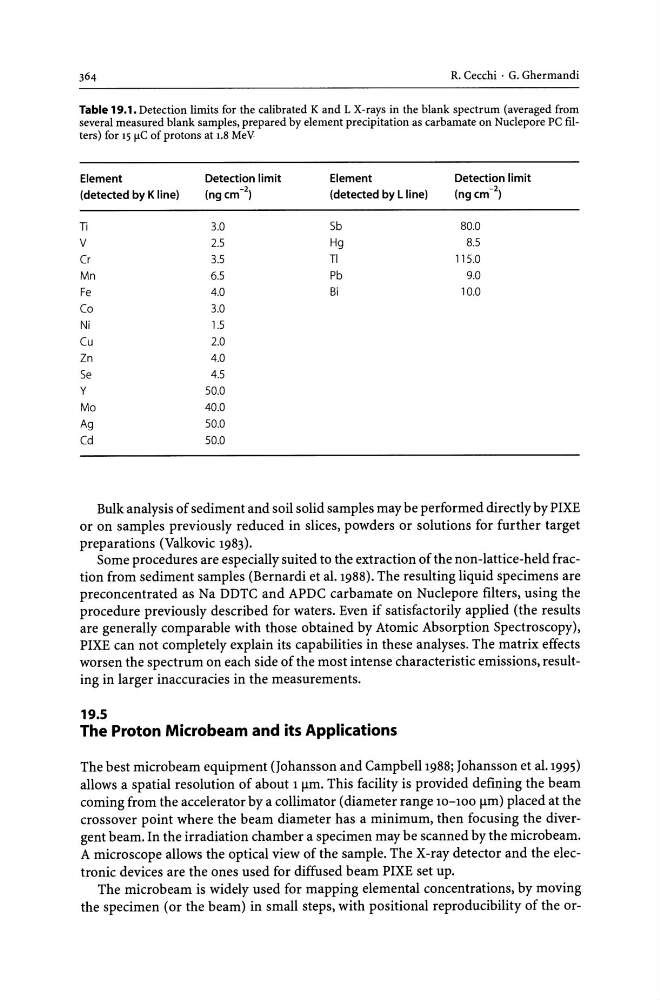
Table 19.1. Detection limits for the calibrated K and L X-rays in the blank spectrum (averaged from
several measured blank samples, prepared by element precipitation as carbamate on Nuclepore PC filters) for 15 flC of protons at 1.8 MeV
Element
Detection limit
Element
Detection limit
(detected by Kline)
(ngcm- 2 )
(detected by L line)
(ngcm- 2 )
Ti
3.0
Sb
80.0
V
2.5
Hg
8.5
Cr
3.5
TI
115.0
Mn
6.5
Pb
9.0
Fe
4.0
Bi
10.0
Co
3.0
Ni
1.5
Cu
2.0
Zn
4.0
Se
4.5
y
50.0
Mo
40.0
Ag
50.0
Cd
50.0
Bulk analysis of sediment and soil solid samples may be performed directly by PIXE
or on samples previously reduced in slices, powders or solutions for further target
preparations (Valkovic 1983).
Some procedures are especially suited to the extraction of the non-lattice-held fraction from sediment samples (Bernardi et al. 1988). The resulting liquid specimens are
preconcentrated as Na DDTC and APDC carbamate on Nuclepore filters, using the
procedure previously described for waters. Even if satisfactorily applied (the results
are generally comparable with those obtained by Atomic Absorption Spectroscopy),
PIXE can not completely explain its capabilities in these analyses. The matrix effects
worsen the spectrum on each side of the most intense characteristic emissions, resulting in larger inaccuracies in the measurements.
19.5
The Proton Microbeam and its Applications
The best microbeam equipment (Johansson and Campbell 1988; Johansson et al. 1995)
allows a spatial resolution of about 1 flm. This facility is provided defining the beam
coming from the accelerator by a collimator (diameter range 10-100 flm) placed at the
crossover point where the beam diameter has a minimum, then focusing the divergent beam. In the irradiation chamber a specimen may be scanned by the microbeam.
A microscope allows the optical view of the sample. The X-ray detector and the electronic devices are the ones used for diffused beam PIXE set up.
The microbe am is widely used for mapping elemental concentrations, by moving
the specimen (or the beam) in small steps, with positional reproducibility of the or-
Table 19.1. Detection limits for the calibrated K and L X-rays in the blank spectrum (averaged from
several measured blank samples, prepared by element precipitation as carbamate on Nuclepore PC filters) for 15 flC of protons at 1.8 MeV
Element
Detection limit
Element
Detection limit
(detected by Kline)
(ngcm- 2 )
(detected by L line)
(ngcm- 2 )
Ti
3.0
Sb
80.0
V
2.5
Hg
8.5
Cr
3.5
TI
115.0
Mn
6.5
Pb
9.0
Fe
4.0
Bi
10.0
Co
3.0
Ni
1.5
Cu
2.0
Zn
4.0
Se
4.5
y
50.0
Mo
40.0
Ag
50.0
Cd
50.0
Bulk analysis of sediment and soil solid samples may be performed directly by PIXE
or on samples previously reduced in slices, powders or solutions for further target
preparations (Valkovic 1983).
Some procedures are especially suited to the extraction of the non-lattice-held fraction from sediment samples (Bernardi et al. 1988). The resulting liquid specimens are
preconcentrated as Na DDTC and APDC carbamate on Nuclepore filters, using the
procedure previously described for waters. Even if satisfactorily applied (the results
are generally comparable with those obtained by Atomic Absorption Spectroscopy),
PIXE can not completely explain its capabilities in these analyses. The matrix effects
worsen the spectrum on each side of the most intense characteristic emissions, resulting in larger inaccuracies in the measurements.
19.5
The Proton Microbeam and its Applications
The best microbeam equipment (Johansson and Campbell 1988; Johansson et al. 1995)
allows a spatial resolution of about 1 flm. This facility is provided defining the beam
coming from the accelerator by a collimator (diameter range 10-100 flm) placed at the
crossover point where the beam diameter has a minimum, then focusing the divergent beam. In the irradiation chamber a specimen may be scanned by the microbeam.
A microscope allows the optical view of the sample. The X-ray detector and the electronic devices are the ones used for diffused beam PIXE set up.
The microbe am is widely used for mapping elemental concentrations, by moving
the specimen (or the beam) in small steps, with positional reproducibility of the or-