

M.E. Farago
Communities who eat large amounts of contaminated seafood are at risk from neurological damage and harm to the unborn foetus. The effects of methyl mercury on
humans and dose response relationships have been discussed (WHO/IPCS 1990). The
biomarkers of exposure are blood and hair for methyl mercury and urine for inorganic
mercury. Mean reference values are (WHO/IPCS 1990): blood 8 flg rl; hair, 2 flg g-I; urine
4 flg rl. Although hair is the preferred indicator, the concentrations in hair can be affected by a number of factors (Suzuki 1988). Until recently HgT concentrations in the
hair of pregnant women up to 10-20 flg g-I have been considered to be safe (WHOI
IPCS 1990), however recent data suggests that this value may be too high (Grandjean
et al. 1997, 1998).
Methyl mercury is well absorbed and the biological half life in fish has been reported to be in the range 300-1000 days, and that for inorganic mercury of about
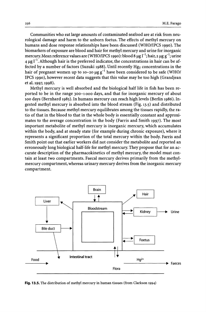
100 days (Bernhard 1985). In humans mercury can reach high levels (Berlin 1986). Ingested methyl mercury is absorbed into the blood stream (Fig. 13.5) and distributed
to the tissues. Because methyl mercury equilibrates among the tissues rapidly, the ratio of that in the blood to that in the whole body is essentially constant and approximates to the average concentration in the body (Farris and Smith 1997). The most
important metabolite of methyl mercury is inorganic mercury, which accumulates
within the body, and at steady state (for example during chronic exposure), where it
represents a significant proportion of the total mercury within the body. Farris and
Smith point out that earlier workers did not consider the metabolite and reported an
erroneously long biological half-life for methyl mercury. They propose that for an accurate description of the pharmacokinetics of methyl mercury, the model must contain at least two compartments. Faecal mercury derives primarily from the methylmercury compartment, whereas urinary mercury derives from the inorganic mercury
compartment.
Brain
/1
Hair
Liver
Bloodstream
I
~
Kidney
Urine
;:=:!I Foetus
Food
Intestinal tract
Hg2+
----------+~ Faeces
Flora
Fig. 13.5. The distribution of methyl mercury in human tissues (from Clarkson 1994)
Communities who eat large amounts of contaminated seafood are at risk from neurological damage and harm to the unborn foetus. The effects of methyl mercury on
humans and dose response relationships have been discussed (WHO/IPCS 1990). The
biomarkers of exposure are blood and hair for methyl mercury and urine for inorganic
mercury. Mean reference values are (WHO/IPCS 1990): blood 8 flg rl; hair, 2 flg g-I; urine
4 flg rl. Although hair is the preferred indicator, the concentrations in hair can be affected by a number of factors (Suzuki 1988). Until recently HgT concentrations in the
hair of pregnant women up to 10-20 flg g-I have been considered to be safe (WHOI
IPCS 1990), however recent data suggests that this value may be too high (Grandjean
et al. 1997, 1998).
Methyl mercury is well absorbed and the biological half life in fish has been reported to be in the range 300-1000 days, and that for inorganic mercury of about
100 days (Bernhard 1985). In humans mercury can reach high levels (Berlin 1986). Ingested methyl mercury is absorbed into the blood stream (Fig. 13.5) and distributed
to the tissues. Because methyl mercury equilibrates among the tissues rapidly, the ratio of that in the blood to that in the whole body is essentially constant and approximates to the average concentration in the body (Farris and Smith 1997). The most
important metabolite of methyl mercury is inorganic mercury, which accumulates
within the body, and at steady state (for example during chronic exposure), where it
represents a significant proportion of the total mercury within the body. Farris and
Smith point out that earlier workers did not consider the metabolite and reported an
erroneously long biological half-life for methyl mercury. They propose that for an accurate description of the pharmacokinetics of methyl mercury, the model must contain at least two compartments. Faecal mercury derives primarily from the methylmercury compartment, whereas urinary mercury derives from the inorganic mercury
compartment.
Brain
/1
Hair
Liver
Bloodstream
I
~
Kidney
Urine
;:=:!I Foetus
Food
Intestinal tract
Hg2+
----------+~ Faeces
Flora
Fig. 13.5. The distribution of methyl mercury in human tissues (from Clarkson 1994)