

416
M.Mascini
The electrodes modified by calf thymus ssDNA immobilization were examined in
0.2 M acetate buffer pH 5.0, using an initial potential of +0.5 V and a constant current of
+6 flA. The guanine peak area following baseline fitting was used as the analytical signal.
18.2.2
DNA Sensor for Binding Compounds with an Affinity for DNA
The procedure consisted of the following steps: calf thymus DNA immobilization on
the electrode surface, dipping the electrode in the sample/blank solution, and electrochemical interrogation of the surface.
Calf thymus immobilization consisted of an electrochemical oxidation (+1.6 V VS. SCE
for 1 min in 0.2 M acetate buffer pH 5.0); then the electrode was immersed in a stirred
buffer solution containing 10-20 mg rl of single stranded or double stranded calf thymus DNA. This immobilization step lasted for 2 min, holding the electrode surface at
a potential of +0.5 V vs. SCE. The electrode was then washed with buffer solution for
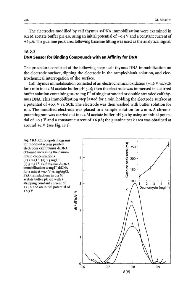
10 s. The modified electrode was placed in a sample solution for 2 min. A chronopotentiogram was carried out in 0.2 M acetate buffer pH 5.0 by using an initial potential of +0.5 V and a constant current of +6 flA; the guanine peak area was obtained at
around +1 V (see Fig. 18.1).
Fig. 18.1. Chronopotentiograms
for modified screen printed
electrodes calf thymus dsDNA
obtained increasing the daunomycin concentrations
(a) 1 mg rl, (b) 2.5 mg rl,
(e) 5 mgrl. Calf thymus dsDNA
immobilization: 10 mg r' dsDNA
for 2 min at +0.5 V vs. AgI AgCI.
PSA transduction: in 0.2 M
acetate buffer pH 5.0 with a
stripping constant current of
+1 f.LA and an initial potential of
+0.5V
4
3
~
~
~
.....
.1Q 2
0.7
"VI
.§. 250
~
ftI
~ 200
~
0.8
E(V)
0.9
M.Mascini
The electrodes modified by calf thymus ssDNA immobilization were examined in
0.2 M acetate buffer pH 5.0, using an initial potential of +0.5 V and a constant current of
+6 flA. The guanine peak area following baseline fitting was used as the analytical signal.
18.2.2
DNA Sensor for Binding Compounds with an Affinity for DNA
The procedure consisted of the following steps: calf thymus DNA immobilization on
the electrode surface, dipping the electrode in the sample/blank solution, and electrochemical interrogation of the surface.
Calf thymus immobilization consisted of an electrochemical oxidation (+1.6 V VS. SCE
for 1 min in 0.2 M acetate buffer pH 5.0); then the electrode was immersed in a stirred
buffer solution containing 10-20 mg rl of single stranded or double stranded calf thymus DNA. This immobilization step lasted for 2 min, holding the electrode surface at
a potential of +0.5 V vs. SCE. The electrode was then washed with buffer solution for
10 s. The modified electrode was placed in a sample solution for 2 min. A chronopotentiogram was carried out in 0.2 M acetate buffer pH 5.0 by using an initial potential of +0.5 V and a constant current of +6 flA; the guanine peak area was obtained at
around +1 V (see Fig. 18.1).
Fig. 18.1. Chronopotentiograms
for modified screen printed
electrodes calf thymus dsDNA
obtained increasing the daunomycin concentrations
(a) 1 mg rl, (b) 2.5 mg rl,
(e) 5 mgrl. Calf thymus dsDNA
immobilization: 10 mg r' dsDNA
for 2 min at +0.5 V vs. AgI AgCI.
PSA transduction: in 0.2 M
acetate buffer pH 5.0 with a
stripping constant current of
+1 f.LA and an initial potential of
+0.5V
4
3
~
~
~
.....
.1Q 2
0.7
"VI
.§. 250
~
ftI
~ 200
~
0.8
E(V)
0.9