

81
Chapter four: Pressure regulation
bubble formation was unable to detect bubbles (Houser et al. 2010). In contrast, a low level of
bubble incidence has been detected in stranded (common and white-sided) dolphins using
B-mode ultrasound. Furthermore, these dolphins showed normal behavior upon release
and did not restrand, suggesting some tolerance to bubble formation (Dennison et al. 2012).
Bubbles have also been observed from marine mammals bycaught in fishing nets, which died
at depth (Moore et al. 2009). These bubbles suggest the animals’ tissues were supersaturated
sufficiently to cause bubble formation when depressurized (as nets were hauled). However,
whether tissue and blood N 2 levels represented the routine load at the time of entrapment or
whether these levels were elevated as animals struggled in the nets is not clear. Two recent
studies have reported neural deficiencies in California sea lions admitted to a rehabilitation
facility (Van Bonn et al. 2011, 2013). In both instances, cerebral gas lesions were observed following magnetic resonance imaging (MRI). While the etiology cannot be determined, it was
postulated that barotrauma would be one plausible explanation.
These cases highlight a growing body of evidence that indicates that our understanding how marine vertebrates manage gases during diving is very rudimentary, and that
they may often experience blood and tissue tensions that are higher than previously
thought (Hooker et al. 2012), and that may cause bubbles to form (de Quiros et al. 2013b;
Dennison et al. 2012; Garcia-Parraga et al. 2014; Moore et al. 2009; Moore and Early 2004).
4.2.6.4 Shallow-water blackout
While shallow-water blackout has not been observed in marine mammals, it has been
documented for human breath-hold divers and, therefore, would be expected to be a
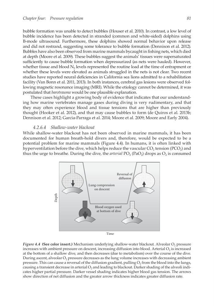
potential problem for marine mammals (Figure 4.4). In humans, it is often linked with
hyperventilation before the dive, which helps reduce the vascular CO 2 tension (PCO 2 ) and
thus the urge to breathe. During the dive, the arterial PO 2 (PaO 2 ) drops as O 2 is consumed
Reversal of
diffusion
Blood oxygen used
at bottom of dive
Lung compression
on descent
Depth
Time
Figure 4.4 (See color insert.) Mechanism underlying shallow-water blackout. Alveolar O 2 pressure
increases with ambient pressure on descent, increasing diffusion into blood. Arterial O 2 is increased
at the bottom of a shallow dive, and then decreases (due to metabolism) over the course of the dive.
During ascent, alveolar O 2 pressure decreases as the lung volume increases with decreasing ambient
pressure. This can cause a reversal of the diffusion gradient, pulling O 2 from the blood into the lungs,
causing a transient decrease in arterial O 2 and leading to blackout. Darker shading of the alveoli indicates higher partial pressure. Darker vessel shading indicates higher blood gas tension. The arrows
show direction of net diffusion and the greater arrow thickness indicates greater diffusion rate.
Chapter four: Pressure regulation
bubble formation was unable to detect bubbles (Houser et al. 2010). In contrast, a low level of
bubble incidence has been detected in stranded (common and white-sided) dolphins using
B-mode ultrasound. Furthermore, these dolphins showed normal behavior upon release
and did not restrand, suggesting some tolerance to bubble formation (Dennison et al. 2012).
Bubbles have also been observed from marine mammals bycaught in fishing nets, which died
at depth (Moore et al. 2009). These bubbles suggest the animals’ tissues were supersaturated
sufficiently to cause bubble formation when depressurized (as nets were hauled). However,
whether tissue and blood N 2 levels represented the routine load at the time of entrapment or
whether these levels were elevated as animals struggled in the nets is not clear. Two recent
studies have reported neural deficiencies in California sea lions admitted to a rehabilitation
facility (Van Bonn et al. 2011, 2013). In both instances, cerebral gas lesions were observed following magnetic resonance imaging (MRI). While the etiology cannot be determined, it was
postulated that barotrauma would be one plausible explanation.
These cases highlight a growing body of evidence that indicates that our understanding how marine vertebrates manage gases during diving is very rudimentary, and that
they may often experience blood and tissue tensions that are higher than previously
thought (Hooker et al. 2012), and that may cause bubbles to form (de Quiros et al. 2013b;
Dennison et al. 2012; Garcia-Parraga et al. 2014; Moore et al. 2009; Moore and Early 2004).
4.2.6.4 Shallow-water blackout
While shallow-water blackout has not been observed in marine mammals, it has been
documented for human breath-hold divers and, therefore, would be expected to be a
potential problem for marine mammals (Figure 4.4). In humans, it is often linked with
hyperventilation before the dive, which helps reduce the vascular CO 2 tension (PCO 2 ) and
thus the urge to breathe. During the dive, the arterial PO 2 (PaO 2 ) drops as O 2 is consumed
Reversal of
diffusion
Blood oxygen used
at bottom of dive
Lung compression
on descent
Depth
Time
Figure 4.4 (See color insert.) Mechanism underlying shallow-water blackout. Alveolar O 2 pressure
increases with ambient pressure on descent, increasing diffusion into blood. Arterial O 2 is increased
at the bottom of a shallow dive, and then decreases (due to metabolism) over the course of the dive.
During ascent, alveolar O 2 pressure decreases as the lung volume increases with decreasing ambient
pressure. This can cause a reversal of the diffusion gradient, pulling O 2 from the blood into the lungs,
causing a transient decrease in arterial O 2 and leading to blackout. Darker shading of the alveoli indicates higher partial pressure. Darker vessel shading indicates higher blood gas tension. The arrows
show direction of net diffusion and the greater arrow thickness indicates greater diffusion rate.