

206
The Biology of Sea Turtles, Vol. II
7.3 HATCHLING GONADS
Attempts have been made to visually distinguish between testis and ovary of
hatchling marine turtles, but the technique has not proven to be useful (Whitmore
et al., 1985; Mrosovsky and Benabib, 1990). At present, only two methods can be
used to reliably determine the sex of a hatchling marine turtle: (1) histological
examination and (2) serological typing (blood sampling) (Merchant-Larios, 1999).
The former requires that the hatchling be killed and the gonads removed and, after
preparation, examined under a microscope. The method is useful in the experimental
context to establish the pivotal temperature for a population under management. The
use of blood does not require the killing of hatchlings, but hormonal analysis (via
radioimmunoassay) is expensive and works best on slightly older post-hatchlings
(curved carapace length [CCL] > 30 cm) (Owens and Ruiz, 1980; Wibbels et al.,
1998) because of the volumes of blood needed. Regardless of the method used, the
pivotal temperature is useful in planning for management of the population and
understanding of the sex ratio of hatchlings leaving the beach (Godfrey et al., 1996).
The gonads of hatchling marine turtles are morphologically defined by the time
the turtles reach the beach surface (Table 7.2). At emergence the gonad appears as
a whitish, elongate structure on the ventral surface of the kidney (Figure 7.2); it
cannot be distinguished by eye as an ovary or a testis.
7.3.1 O VARY
Histologically, the cortex of the ovary is differentiated into columnar shaped cells
with germinal cells spaced among them near the basal membrane (Figure 7.3). The
medulla is dense with occasional small strings of cells among blood vessels and
other cells; no tubules with open lumen are present.
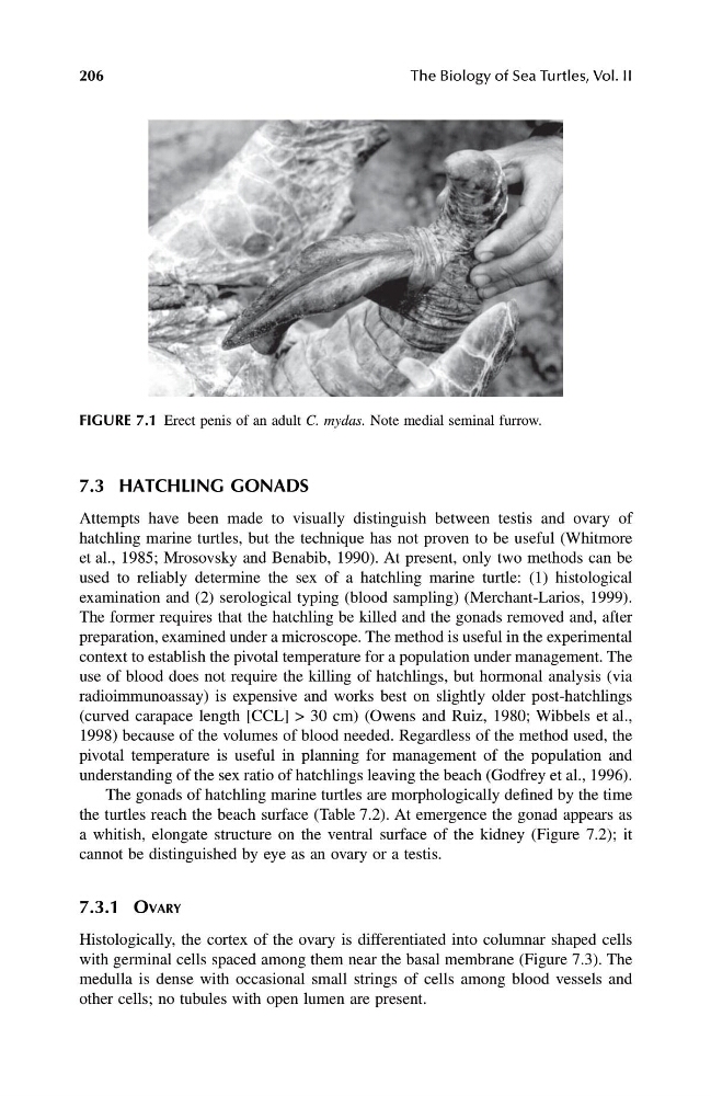
FIGURE 7.1 Erect penis of an adult C. mydas. Note medial seminal furrow.
1123 book.book Page 206 Monday, November 11, 2002 11:11 AM
The Biology of Sea Turtles, Vol. II
7.3 HATCHLING GONADS
Attempts have been made to visually distinguish between testis and ovary of
hatchling marine turtles, but the technique has not proven to be useful (Whitmore
et al., 1985; Mrosovsky and Benabib, 1990). At present, only two methods can be
used to reliably determine the sex of a hatchling marine turtle: (1) histological
examination and (2) serological typing (blood sampling) (Merchant-Larios, 1999).
The former requires that the hatchling be killed and the gonads removed and, after
preparation, examined under a microscope. The method is useful in the experimental
context to establish the pivotal temperature for a population under management. The
use of blood does not require the killing of hatchlings, but hormonal analysis (via
radioimmunoassay) is expensive and works best on slightly older post-hatchlings
(curved carapace length [CCL] > 30 cm) (Owens and Ruiz, 1980; Wibbels et al.,
1998) because of the volumes of blood needed. Regardless of the method used, the
pivotal temperature is useful in planning for management of the population and
understanding of the sex ratio of hatchlings leaving the beach (Godfrey et al., 1996).
The gonads of hatchling marine turtles are morphologically defined by the time
the turtles reach the beach surface (Table 7.2). At emergence the gonad appears as
a whitish, elongate structure on the ventral surface of the kidney (Figure 7.2); it
cannot be distinguished by eye as an ovary or a testis.
7.3.1 O VARY
Histologically, the cortex of the ovary is differentiated into columnar shaped cells
with germinal cells spaced among them near the basal membrane (Figure 7.3). The
medulla is dense with occasional small strings of cells among blood vessels and
other cells; no tubules with open lumen are present.
FIGURE 7.1 Erect penis of an adult C. mydas. Note medial seminal furrow.
1123 book.book Page 206 Monday, November 11, 2002 11:11 AM