

Ontogeny of Marine Turtle Gonads
203
7.2.3 G ONADAL M ORPHOGENESIS
Before sexual differentiation, the structure of the gonadal primordia is similar in
both presumptive sexes. The gonadal primordia has two regions: the cortex (outer),
which is characterized by a single layer of cuboidal epithelial cells with embedded
germinal primordial cells, and the medulla (inner), which is derived from mesenchyme cells within the middle of the undifferentiated gonad. As sexual differentiation
occurs (during stages 24–29, Merchant-Larios et al., 1989), the cells of the gonadal
primordia propagate into two distinct and opposite patterns. At this time, changes
in both the cortex and medulla are visible. Most of the primordial germ cells are
situated in the cortex; both nerve and blood vessels penetrate the mass of mesenchyme cells in the medullary area.
In presumptive females, the ovary results from the simultaneous proliferation of
cells in the cortex and regressive modification in the medulla. The cells of the cortex
become more columnar in shape and the layer becomes thicker; primordial germinal
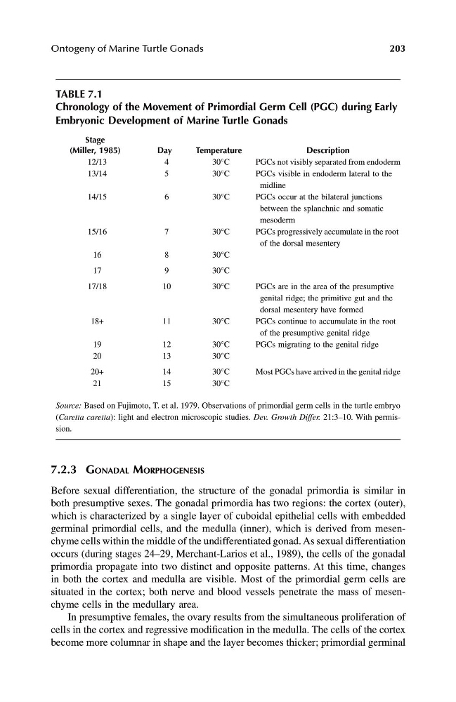
TABLE 7.1
Chronology of the Movement of Primordial Germ Cell (PGC) during Early
Embryonic Development of Marine Turtle Gonads
Stage
(Miller, 1985)
Day
Temperature
Description
12/13
4
30°C
PGCs not visibly separated from endoderm
13/14
5
30°C
PGCs visible in endoderm lateral to the
midline
14/15
6
30°C
PGCs occur at the bilateral junctions
between the splanchnic and somatic
mesoderm
15/16
7
30°C
PGCs progressively accumulate in the root
of the dorsal mesentery
16
8
30°C
17
9
30°C
17/18
10
30°C
PGCs are in the area of the presumptive
genital ridge; the primitive gut and the
dorsal mesentery have formed
18+
11
30°C
PGCs continue to accumulate in the root
of the presumptive genital ridge
19
12
30°C
PGCs migrating to the genital ridge
20
13
30°C
20+
14
30°C
Most PGCs have arrived in the genital ridge
21
15
30°C
Source: Based on Fujimoto, T. et al. 1979. Observations of primordial germ cells in the turtle embryo
( Caretta caretta ): light and electron microscopic studies. Dev. Growth Differ. 21:3–10. With permission.
1123 book.book Page 203 Monday, November 11, 2002 11:11 AM
203
7.2.3 G ONADAL M ORPHOGENESIS
Before sexual differentiation, the structure of the gonadal primordia is similar in
both presumptive sexes. The gonadal primordia has two regions: the cortex (outer),
which is characterized by a single layer of cuboidal epithelial cells with embedded
germinal primordial cells, and the medulla (inner), which is derived from mesenchyme cells within the middle of the undifferentiated gonad. As sexual differentiation
occurs (during stages 24–29, Merchant-Larios et al., 1989), the cells of the gonadal
primordia propagate into two distinct and opposite patterns. At this time, changes
in both the cortex and medulla are visible. Most of the primordial germ cells are
situated in the cortex; both nerve and blood vessels penetrate the mass of mesenchyme cells in the medullary area.
In presumptive females, the ovary results from the simultaneous proliferation of
cells in the cortex and regressive modification in the medulla. The cells of the cortex
become more columnar in shape and the layer becomes thicker; primordial germinal
TABLE 7.1
Chronology of the Movement of Primordial Germ Cell (PGC) during Early
Embryonic Development of Marine Turtle Gonads
Stage
(Miller, 1985)
Day
Temperature
Description
12/13
4
30°C
PGCs not visibly separated from endoderm
13/14
5
30°C
PGCs visible in endoderm lateral to the
midline
14/15
6
30°C
PGCs occur at the bilateral junctions
between the splanchnic and somatic
mesoderm
15/16
7
30°C
PGCs progressively accumulate in the root
of the dorsal mesentery
16
8
30°C
17
9
30°C
17/18
10
30°C
PGCs are in the area of the presumptive
genital ridge; the primitive gut and the
dorsal mesentery have formed
18+
11
30°C
PGCs continue to accumulate in the root
of the presumptive genital ridge
19
12
30°C
PGCs migrating to the genital ridge
20
13
30°C
20+
14
30°C
Most PGCs have arrived in the genital ridge
21
15
30°C
Source: Based on Fujimoto, T. et al. 1979. Observations of primordial germ cells in the turtle embryo
( Caretta caretta ): light and electron microscopic studies. Dev. Growth Differ. 21:3–10. With permission.
1123 book.book Page 203 Monday, November 11, 2002 11:11 AM