

Reproductive Cycles of Males and Females
143
5.5 FEMALES
5.5.1 ANATOMY OF THE FEMALE REPRODUCTIVE SYSTEM
Female sea turtles have paired reproductive organs located abdominally. During
puberty, hormonal changes increase the size and structure of both the ovary and
oviduct. In comparison with immature or pubescent females, mature females typically have an ovary with an expanded stroma and a convoluted oviduct at least 1.5
cm in diameter (adjacent to the ovary) suspended in the body cavity. Oviducts of
adults are very long, and lengths of 4–5 and >6 m have been recorded from L.
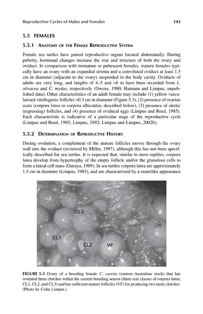
olivacea and C. mydas, respectively (Owens, 1980; Hamann and Limpus, unpublished data). Other characteristics of an adult female may include (1) yellow vascularized vitellogenic follicles >0.3 cm in diameter (Figure 5.3), (2) presence of ovarian
scars (corpora lutea or corpora albicantia; described below), (3) presence of atretic
(regressing) follicles, and (4) presence of oviducal eggs (Limpus and Reed, 1985).
Each characteristic is indicative of a particular stage of the reproductive cycle
(Limpus and Reed, 1985; Limpus, 1992; Limpus and Limpus, 2002b).
5.5.2 DETERMINATION OF REPRODUCTIVE HISTORY
During ovulation, a complement of the mature follicles moves through the ovary
wall into the oviduct (reviewed by Miller, 1997), although this has not been specifically described for sea turtles. It is expected that, similar to most reptiles, corpora
lutea develop from hypertrophy of the empty follicle and/or the granulosa cells to
form a luteal cell mass (Guraya, 1989). In sea turtles corpora lutea are approximately
1.5 cm in diameter (Limpus, 1985), and are characterized by a craterlike appearance
FIGURE 5.3 Ovary of a breeding female C. caretta (eastern Australian stock) that has
ovulated three clutches within the current breeding season (three size classes of corpora lutea;
CL1, CL2, and CL3) and has sufficient mature follicles (VF) for producing two more clutches.
(Photo by Colin Limpus.)
1123 book.book Page 143 Thursday, November 14, 2002 11:50 AM
143
5.5 FEMALES
5.5.1 ANATOMY OF THE FEMALE REPRODUCTIVE SYSTEM
Female sea turtles have paired reproductive organs located abdominally. During
puberty, hormonal changes increase the size and structure of both the ovary and
oviduct. In comparison with immature or pubescent females, mature females typically have an ovary with an expanded stroma and a convoluted oviduct at least 1.5
cm in diameter (adjacent to the ovary) suspended in the body cavity. Oviducts of
adults are very long, and lengths of 4–5 and >6 m have been recorded from L.
olivacea and C. mydas, respectively (Owens, 1980; Hamann and Limpus, unpublished data). Other characteristics of an adult female may include (1) yellow vascularized vitellogenic follicles >0.3 cm in diameter (Figure 5.3), (2) presence of ovarian
scars (corpora lutea or corpora albicantia; described below), (3) presence of atretic
(regressing) follicles, and (4) presence of oviducal eggs (Limpus and Reed, 1985).
Each characteristic is indicative of a particular stage of the reproductive cycle
(Limpus and Reed, 1985; Limpus, 1992; Limpus and Limpus, 2002b).
5.5.2 DETERMINATION OF REPRODUCTIVE HISTORY
During ovulation, a complement of the mature follicles moves through the ovary
wall into the oviduct (reviewed by Miller, 1997), although this has not been specifically described for sea turtles. It is expected that, similar to most reptiles, corpora
lutea develop from hypertrophy of the empty follicle and/or the granulosa cells to
form a luteal cell mass (Guraya, 1989). In sea turtles corpora lutea are approximately
1.5 cm in diameter (Limpus, 1985), and are characterized by a craterlike appearance
FIGURE 5.3 Ovary of a breeding female C. caretta (eastern Australian stock) that has
ovulated three clutches within the current breeding season (three size classes of corpora lutea;
CL1, CL2, and CL3) and has sufficient mature follicles (VF) for producing two more clutches.
(Photo by Colin Limpus.)
1123 book.book Page 143 Thursday, November 14, 2002 11:50 AM