

138
The Biology of Sea Turtles, Vol. II
Similarly, ultrasonography (see Rostal et al. , 1990; Owens, 1999; Wibbels et al. ,
2000) has been used extensively in several sea turtle projects for quantitative analysis
of follicle size, examination of intraoviducal egg development, characterization of
reproductive condition, prediction of the likelihood of future reproductive events in
breeding females, and the assessment of reproductive condition for tracking studies
(Rostal et al ., 1990; 1996; 1997; 1998; 2001; Plotkin et al ., 1995). However, this
noninvasive procedure is limited by its inability to image oviducts and ovarian
features such as corpora lutea and corpora albicantia. Thus, it cannot be used to
quantify reproductive maturity in nonbreeding females or past breeding history in
adult females. In addition, its use is currently restricted to the examination of
breeding females, and continuing work (unpublished) by both Owens and Limpus
has shown that they were unable to obtain recognizable images of ovaries in nonbreeding females, or of testes or epididymis using ultrasonography. Regardless, the
development of this technique over the last decade has been significant, and its usage
promises to further enhance our understanding of the reproductive biology of adult
female sea turtles.
5.4 MALES
5.4.1 A NATOMY OF THE M ALE R EPRODUCTIVE S YSTEM
Similar to most vertebrates, the male reproductive system in sea turtles is composed
of simultaneously functioning paired testes and associated ducts (ducts epididymis,
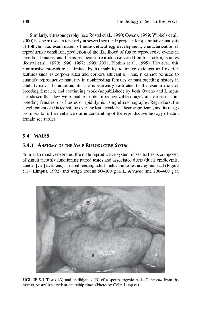
ductus [vas] deferens). In nonbreeding adult males the testes are cylindrical (Figure
5.1) (Limpus, 1992) and weigh around 50–100 g in L. olivacea and 200–400 g in
FIGURE 5.1 Testis (A) and epididymus (B) of a spermatogenic male C. caretta from the
eastern Australian stock at courtship time. (Photo by Colin Limpus.)
1123 book.book Page 138 Thursday, November 14, 2002 11:50 AM
The Biology of Sea Turtles, Vol. II
Similarly, ultrasonography (see Rostal et al. , 1990; Owens, 1999; Wibbels et al. ,
2000) has been used extensively in several sea turtle projects for quantitative analysis
of follicle size, examination of intraoviducal egg development, characterization of
reproductive condition, prediction of the likelihood of future reproductive events in
breeding females, and the assessment of reproductive condition for tracking studies
(Rostal et al ., 1990; 1996; 1997; 1998; 2001; Plotkin et al ., 1995). However, this
noninvasive procedure is limited by its inability to image oviducts and ovarian
features such as corpora lutea and corpora albicantia. Thus, it cannot be used to
quantify reproductive maturity in nonbreeding females or past breeding history in
adult females. In addition, its use is currently restricted to the examination of
breeding females, and continuing work (unpublished) by both Owens and Limpus
has shown that they were unable to obtain recognizable images of ovaries in nonbreeding females, or of testes or epididymis using ultrasonography. Regardless, the
development of this technique over the last decade has been significant, and its usage
promises to further enhance our understanding of the reproductive biology of adult
female sea turtles.
5.4 MALES
5.4.1 A NATOMY OF THE M ALE R EPRODUCTIVE S YSTEM
Similar to most vertebrates, the male reproductive system in sea turtles is composed
of simultaneously functioning paired testes and associated ducts (ducts epididymis,
ductus [vas] deferens). In nonbreeding adult males the testes are cylindrical (Figure
5.1) (Limpus, 1992) and weigh around 50–100 g in L. olivacea and 200–400 g in
FIGURE 5.1 Testis (A) and epididymus (B) of a spermatogenic male C. caretta from the
eastern Australian stock at courtship time. (Photo by Colin Limpus.)
1123 book.book Page 138 Thursday, November 14, 2002 11:50 AM