

Sensory Biology of Sea Turtles
81
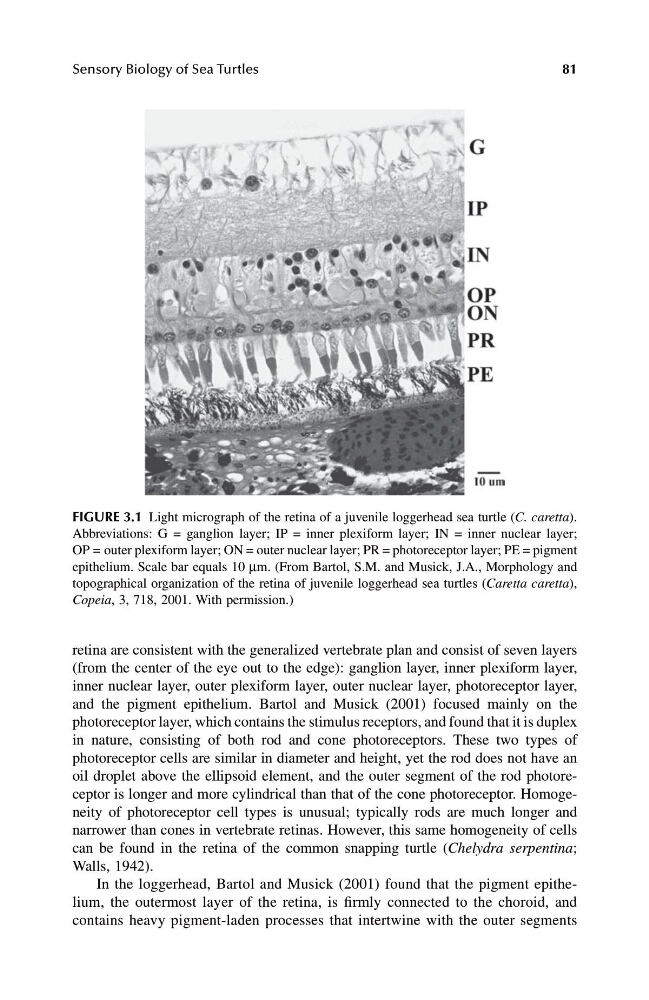
retina are consistent with the generalized vertebrate plan and consist of seven layers
(from the center of the eye out to the edge): ganglion layer, inner plexiform layer,
inner nuclear layer, outer plexiform layer, outer nuclear layer, photoreceptor layer,
and the pigment epithelium. Bartol and Musick (2001) focused mainly on the
photoreceptor layer, which contains the stimulus receptors, and found that it is duplex
in nature, consisting of both rod and cone photoreceptors. These two types of
photoreceptor cells are similar in diameter and height, yet the rod does not have an
oil droplet above the ellipsoid element, and the outer segment of the rod photoreceptor is longer and more cylindrical than that of the cone photoreceptor. Homogeneity of photoreceptor cell types is unusual; typically rods are much longer and
narrower than cones in vertebrate retinas. However, this same homogeneity of cells
can be found in the retina of the common snapping turtle ( Chelydra serpentina ;
Walls, 1942).
In the loggerhead, Bartol and Musick (2001) found that the pigment epithelium, the outermost layer of the retina, is firmly connected to the choroid, and
contains heavy pigment-laden processes that intertwine with the outer segments
FIGURE 3.1 Light micrograph of the retina of a juvenile loggerhead sea turtle ( C. caretta ).
Abbreviations: G = ganglion layer; IP = inner plexiform layer; IN = inner nuclear layer;
OP = outer plexiform layer; ON = outer nuclear layer; PR = photoreceptor layer; PE = pigment
epithelium. Scale bar equals 10 m m. (From Bartol, S.M. and Musick, J.A., Morphology and
topographical organization of the retina of juvenile loggerhead sea turtles ( Caretta caretta ),
Copeia , 3, 718, 2001. With permission.)
1123 book.book Page 81 Monday, November 11, 2002 11:11 AM
81
retina are consistent with the generalized vertebrate plan and consist of seven layers
(from the center of the eye out to the edge): ganglion layer, inner plexiform layer,
inner nuclear layer, outer plexiform layer, outer nuclear layer, photoreceptor layer,
and the pigment epithelium. Bartol and Musick (2001) focused mainly on the
photoreceptor layer, which contains the stimulus receptors, and found that it is duplex
in nature, consisting of both rod and cone photoreceptors. These two types of
photoreceptor cells are similar in diameter and height, yet the rod does not have an
oil droplet above the ellipsoid element, and the outer segment of the rod photoreceptor is longer and more cylindrical than that of the cone photoreceptor. Homogeneity of photoreceptor cell types is unusual; typically rods are much longer and
narrower than cones in vertebrate retinas. However, this same homogeneity of cells
can be found in the retina of the common snapping turtle ( Chelydra serpentina ;
Walls, 1942).
In the loggerhead, Bartol and Musick (2001) found that the pigment epithelium, the outermost layer of the retina, is firmly connected to the choroid, and
contains heavy pigment-laden processes that intertwine with the outer segments
FIGURE 3.1 Light micrograph of the retina of a juvenile loggerhead sea turtle ( C. caretta ).
Abbreviations: G = ganglion layer; IP = inner plexiform layer; IN = inner nuclear layer;
OP = outer plexiform layer; ON = outer nuclear layer; PR = photoreceptor layer; PE = pigment
epithelium. Scale bar equals 10 m m. (From Bartol, S.M. and Musick, J.A., Morphology and
topographical organization of the retina of juvenile loggerhead sea turtles ( Caretta caretta ),
Copeia , 3, 718, 2001. With permission.)
1123 book.book Page 81 Monday, November 11, 2002 11:11 AM