

54
Biology of Sea Turtles, Vol. II
The forelimb is composed of the humerus, radius, ulna, carpals, metacarpals,
and five phalanges (Figure 2.13). The radius and ulna are short in sea turtles and,
in adults, functionally fused by fibrous connective tissue. The flipper blade is formed
by widening and flattening of the wrist bones (radiale, ulnare, centrale, pisiform,
distal carpals) and elongation of the digits (metacarpals and phalanges) (Figure 2.13).
The cheloniid humerus (Figure 2.15) articulates with the shoulder at the glenoid
fossa (Figure 2.14); it is flattened with its head offset from the bone’s shaft (Walker,
1973). A large medial process extends proximally beyond the humeral head and is
a major attachment site for flipper abductor and extensor muscles. Distal to the head
and almost diagonally opposite is the lateral process (deltoid crest), to which attach
flipper protractor muscles. In Dermochelys, the humerus is extremely flattened, with
large portions of the humeral head, distal condyles, and medial process formed by
highly vascular articular cartilages (Figure 2.16).
The Dermochelyidae and Cheloniidae diverge in the types of bone and cartilage
that form their appendicular skeleton. Extensive vascular channels in the cartilage
ends of leatherback long bones (Figure 2.16) are indicative of chondro-osseus bone
formation (Rhodin, 1985; Rhodin et al., 1981). This is unlike cheloniid bone, which
is formed by deposition of relatively thick layers (lamellae) of cortical bone around
a cellular bony core (cancellous bone). These two bone formation types are illustrated
comparatively in gross form by Wyneken (2001) and histologically by Rhodin (1985).
The pelvis is composed of three paired bones: the pubis, ischium, and ilium. The
two ilia are oriented dorsoventrally and articulate with the sacral vertebrae. The ischia,
pubic bones, and cartilages form the largely flat ventral surface of the pelvic girdle. All
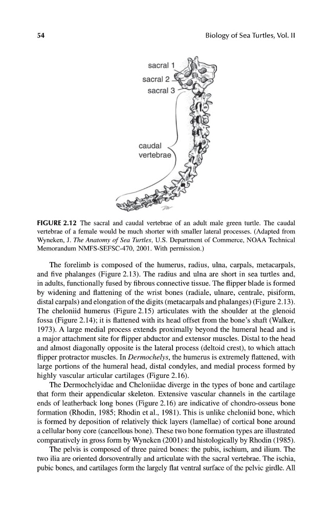
FIGURE 2.12 The sacral and caudal vertebrae of an adult male green turtle. The caudal
vertebrae of a female would be much shorter with smaller lateral processes. (Adapted from
Wyneken, J. The Anatomy of Sea Turtles, U.S. Department of Commerce, NOAA Technical
Memorandum NMFS-SEFSC-470, 2001. With permission.)
1123_C02.fm Page 54 Friday, November 15, 2002 11:16 AM
Biology of Sea Turtles, Vol. II
The forelimb is composed of the humerus, radius, ulna, carpals, metacarpals,
and five phalanges (Figure 2.13). The radius and ulna are short in sea turtles and,
in adults, functionally fused by fibrous connective tissue. The flipper blade is formed
by widening and flattening of the wrist bones (radiale, ulnare, centrale, pisiform,
distal carpals) and elongation of the digits (metacarpals and phalanges) (Figure 2.13).
The cheloniid humerus (Figure 2.15) articulates with the shoulder at the glenoid
fossa (Figure 2.14); it is flattened with its head offset from the bone’s shaft (Walker,
1973). A large medial process extends proximally beyond the humeral head and is
a major attachment site for flipper abductor and extensor muscles. Distal to the head
and almost diagonally opposite is the lateral process (deltoid crest), to which attach
flipper protractor muscles. In Dermochelys, the humerus is extremely flattened, with
large portions of the humeral head, distal condyles, and medial process formed by
highly vascular articular cartilages (Figure 2.16).
The Dermochelyidae and Cheloniidae diverge in the types of bone and cartilage
that form their appendicular skeleton. Extensive vascular channels in the cartilage
ends of leatherback long bones (Figure 2.16) are indicative of chondro-osseus bone
formation (Rhodin, 1985; Rhodin et al., 1981). This is unlike cheloniid bone, which
is formed by deposition of relatively thick layers (lamellae) of cortical bone around
a cellular bony core (cancellous bone). These two bone formation types are illustrated
comparatively in gross form by Wyneken (2001) and histologically by Rhodin (1985).
The pelvis is composed of three paired bones: the pubis, ischium, and ilium. The
two ilia are oriented dorsoventrally and articulate with the sacral vertebrae. The ischia,
pubic bones, and cartilages form the largely flat ventral surface of the pelvic girdle. All
FIGURE 2.12 The sacral and caudal vertebrae of an adult male green turtle. The caudal
vertebrae of a female would be much shorter with smaller lateral processes. (Adapted from
Wyneken, J. The Anatomy of Sea Turtles, U.S. Department of Commerce, NOAA Technical
Memorandum NMFS-SEFSC-470, 2001. With permission.)
1123_C02.fm Page 54 Friday, November 15, 2002 11:16 AM