

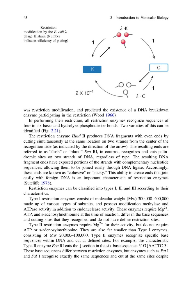
was restriction modification, and predicted the existence of a DNA breakdown
enzyme participating in the restriction (Wood 1966).
In performing their restriction, all restriction enzymes recognize sequences of
four to six bases and hydrolyze phosphodiester bonds. Two varieties of this can be
identified (Fig. 2.21).
The restriction enzyme Hind II produces DNA fragments with even ends by
cutting simultaneously at the same location on two strands from the center of the
recognition side (as indicated by the direction of the arrow). The resulting ends are
referred to as “flush” or “blunt.” Eco RI, in contrast, recognizes and cuts palindromic sites on two strands of DNA, regardless of type. The resulting DNA
fragment ends have exposed portions of the strands with complementary nucleotide
sequences, allowing them to be joined easily through DNA ligase. Accordingly,
these ends are known as “cohesive” or “sticky.” This ability to create ends that join
easily with foreign DNA is an important characteristic of restriction enzymes
(Sutcliffe 1978).
Restriction enzymes can be classified into types I, II, and III according to their
characteristics.
Type I restriction enzymes consist of molecular weight (Mw) 300,000–400,000
made up of various types of subunits, and possess modification methylase and
ATPase activity in addition to endonuclease activity. These enzymes require Mg
2+ ,
ATP, and s-adenosylmethionine at the time of reaction, differ in the base sequences
and cutting sites that they recognize, and do not have define restriction sites.
Type II restriction enzymes require Mg
2+ for their activity, but do not require
ATP or s-adenosylmethionine. They are also far smaller than Type I enzymes,
consisting of Mw 20,000–100,000. Type II enzymes recognize specific base
sequences within DNA and cut at defined sites. For example, the characteristic
Type II enzyme Eco RI cuts the # section in the six-base sequence 5′-G#AATTC-3′.
These base sequences differ between restriction enzymes, but enzymes such as Pst I
and Sal I recognize exactly the same sequences and cut at the same sites despite
Fig. 2.20 Restriction
modification by the E. coli k
phage K strain (Number
indicates efficiency of plating)
48
2 Introduction to Molecular Biology
enzyme participating in the restriction (Wood 1966).
In performing their restriction, all restriction enzymes recognize sequences of
four to six bases and hydrolyze phosphodiester bonds. Two varieties of this can be
identified (Fig. 2.21).
The restriction enzyme Hind II produces DNA fragments with even ends by
cutting simultaneously at the same location on two strands from the center of the
recognition side (as indicated by the direction of the arrow). The resulting ends are
referred to as “flush” or “blunt.” Eco RI, in contrast, recognizes and cuts palindromic sites on two strands of DNA, regardless of type. The resulting DNA
fragment ends have exposed portions of the strands with complementary nucleotide
sequences, allowing them to be joined easily through DNA ligase. Accordingly,
these ends are known as “cohesive” or “sticky.” This ability to create ends that join
easily with foreign DNA is an important characteristic of restriction enzymes
(Sutcliffe 1978).
Restriction enzymes can be classified into types I, II, and III according to their
characteristics.
Type I restriction enzymes consist of molecular weight (Mw) 300,000–400,000
made up of various types of subunits, and possess modification methylase and
ATPase activity in addition to endonuclease activity. These enzymes require Mg
2+ ,
ATP, and s-adenosylmethionine at the time of reaction, differ in the base sequences
and cutting sites that they recognize, and do not have define restriction sites.
Type II restriction enzymes require Mg
2+ for their activity, but do not require
ATP or s-adenosylmethionine. They are also far smaller than Type I enzymes,
consisting of Mw 20,000–100,000. Type II enzymes recognize specific base
sequences within DNA and cut at defined sites. For example, the characteristic
Type II enzyme Eco RI cuts the # section in the six-base sequence 5′-G#AATTC-3′.
These base sequences differ between restriction enzymes, but enzymes such as Pst I
and Sal I recognize exactly the same sequences and cut at the same sites despite
Fig. 2.20 Restriction
modification by the E. coli k
phage K strain (Number
indicates efficiency of plating)
48
2 Introduction to Molecular Biology