

repeating units measuring 0.34 nm between base pairs and 3.4 nm between loops
(Chargaff 2012).
In 1953, Watson and Crick proposed the double helix model for DNA based on
Chargaff’s observations and the X-ray diffraction data. Its characteristics were as
follows:
1. Chains consisting of two strands of polynucleotides twist in opposing directions.
In other words, if one strand followed a 5′ ! 3′ pattern, the other would follow
a 3′ ! 5′ pattern.
2. The two strands’ chain is maintained through hydrogen bonds between adenine
and thymine and between guanine and cytosine.
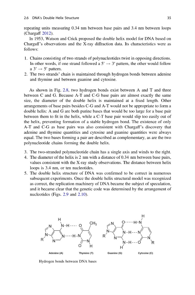
As shown in Fig. 2.8, two hydrogen bonds exist between A and T and three
between C and G. Because A-T and C-G base pairs are almost exactly the same
size, the diameter of the double helix is maintained at a fixed length. Other
arrangements of base pairs besides C-G and A-T would not be appropriate to form a
double helix: A and G are both purine bases that would be too large for a base pair
between them to fit in the helix, while a C-T base pair would slip too easily out of
the helix, preventing formation of a stable hydrogen bond. The existence of only
A-T and C-G as base pairs was also consistent with Chargaff’s discovery that
adenine and thymine quantities and cytosine and guanine quantities were always
equal. The two bases forming a pair are described as complementary, as are the two
polynucleotide chains forming the double helix.
3. The two-stranded polynucleotide chain has a single axis and winds to the right.
4. The diameter of the helix is 2 nm with a distance of 0.34 nm between base pairs,
values consistent with the X-ray study observations. The distance between helix
loops is 3.4 nm, or ten nucleotides.
5. The double helix structure of DNA was confirmed to be correct in numerous
subsequent experiments. Once the double helix structural model was recognized
as correct, the replication machinery of DNA became the subject of speculation,
and it became clear that the genetic code was determined by the arrangement of
nucleotides (Figs. 2.9 and 2.10).
Fig. 2.8 Hydrogen bonds between DNA bases
2.6 DNA’s Double Helix Structure
35
(Chargaff 2012).
In 1953, Watson and Crick proposed the double helix model for DNA based on
Chargaff’s observations and the X-ray diffraction data. Its characteristics were as
follows:
1. Chains consisting of two strands of polynucleotides twist in opposing directions.
In other words, if one strand followed a 5′ ! 3′ pattern, the other would follow
a 3′ ! 5′ pattern.
2. The two strands’ chain is maintained through hydrogen bonds between adenine
and thymine and between guanine and cytosine.
As shown in Fig. 2.8, two hydrogen bonds exist between A and T and three
between C and G. Because A-T and C-G base pairs are almost exactly the same
size, the diameter of the double helix is maintained at a fixed length. Other
arrangements of base pairs besides C-G and A-T would not be appropriate to form a
double helix: A and G are both purine bases that would be too large for a base pair
between them to fit in the helix, while a C-T base pair would slip too easily out of
the helix, preventing formation of a stable hydrogen bond. The existence of only
A-T and C-G as base pairs was also consistent with Chargaff’s discovery that
adenine and thymine quantities and cytosine and guanine quantities were always
equal. The two bases forming a pair are described as complementary, as are the two
polynucleotide chains forming the double helix.
3. The two-stranded polynucleotide chain has a single axis and winds to the right.
4. The diameter of the helix is 2 nm with a distance of 0.34 nm between base pairs,
values consistent with the X-ray study observations. The distance between helix
loops is 3.4 nm, or ten nucleotides.
5. The double helix structure of DNA was confirmed to be correct in numerous
subsequent experiments. Once the double helix structural model was recognized
as correct, the replication machinery of DNA became the subject of speculation,
and it became clear that the genetic code was determined by the arrangement of
nucleotides (Figs. 2.9 and 2.10).
Fig. 2.8 Hydrogen bonds between DNA bases
2.6 DNA’s Double Helix Structure
35