

The Great Barrier Reef
182
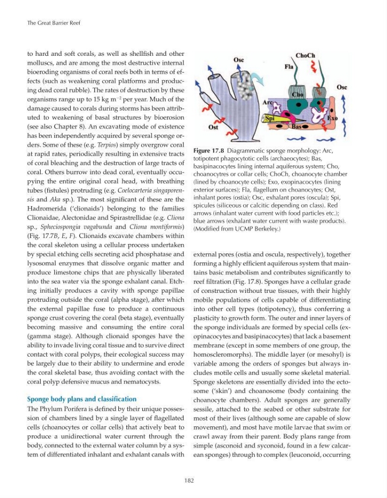
Figure 17.8 Diagrammatic sponge morphology: Arc,
totipotent phagocytotic cells (archaeocytes); Bas,
basipinacocytes lining internal aquiferous system; Cho,
choanocytres or collar cells; ChoCh, choanocyte chamber
(lined by choanocyte cells); Exo, exopinacocytes (lining
exterior surfaces); Fla, flagellum on choanocytes; Ost,
inhalant pores (ostia); Osc, exhalant pores (oscula); Spi,
spicules (siliceous or calcitic depending on class). Red
arrows (inhalant water current with food particles etc.);
blue arrows (exhalant water current with waste products).
(Modified from UCMP Berkeley.)
to hard and soft corals, as well as shellfish and other
molluscs, and are among the most destructive internal
bioeroding organisms of coral reefs both in terms of effects (such as weakening coral platforms and producing dead coral rubble). The rates of destruction by these
organisms range up to 15 kg m
2 per year. Much of the
damage caused to corals during storms has been attributed to weakening of basal structures by bioerosion
(see also Chapter 8). An excavating mode of existence
has been independently acquired by several sponge orders. Some of these (e.g. Terpios) simply overgrow coral
at rapid rates, periodically resulting in extensive tracts
of coral bleaching and the destruction of large tracts of
coral. Others burrow into dead coral, eventually occupying the entire original coral head, with breathing
tubes (fistules) protruding (e.g. Coelocarteria singaporensis and Aka sp.). The most significant of these are the
Hadromerida (‘clionaids’) belonging to the families
Clionaidae, Alectonidae and Spirastrellidae (e.g. Cliona
sp., Spheciospongia vagabunda and Cliona montiformis)
(Fig. 17.7B, E, F). Clionaids excavate chambers within
the coral skeleton using a cellular process undertaken
by special etching cells secreting acid phosphatase and
lysosomal enzymes that dissolve organic matter and
produce limestone chips that are physically liberated
into the sea water via the sponge exhalant canal. Etching initially produces a cavity with sponge papillae
protruding outside the coral (alpha stage), after which
the external papillae fuse to produce a continuous
sponge crust covering the coral (beta stage), eventually
becoming massive and consuming the entire coral
(gamma stage). Although clionaid sponges have the
ability to invade living coral tissue and to survive direct
contact with coral polyps, their ecological success may
be largely due to their ability to undermine and erode
the coral skeletal base, thus avoiding contact with the
coral polyp defensive mucus and nematocysts.
Sponge body plans and classification
The Phylum Porifera is defined by their unique possession of chambers lined by a single layer of flagellated
cells (choanocytes or collar cells) that actively beat to
produce a unidirectional water current through the
body, connected to the external water column by a system of differentiated inhalant and exhalant canals with
external pores (ostia and oscula, respectively), together
forming a highly efficient aquiferous system that maintains basic metabolism and contributes significantly to
reef filtration (Fig. 17.8). Sponges have a cellular grade
of construction without true tissues, with their highly
mobile populations of cells capable of differentiating
into other cell types (totipotency), thus conferring a
plasticity to growth form. The outer and inner layers of
the sponge individuals are formed by special cells (exopinacocytes and basipinacocytes) that lack a basement
membrane (except in some members of one group, the
homoscleromorphs). The middle layer (or mesohyl) is
variable among the orders of sponges but always includes motile cells and usually some skeletal material.
Sponge skeletons are essentially divided into the ectosome (‘skin’) and choanosome (body containing the
choanocyte chambers). Adult sponges are generally
sessile, attached to the seabed or other substrate for
most of their lives (although some are capable of slow
movement), and most have motile larvae that swim or
crawl away from their parent. Body plans range from
simple (asconoid and syconoid, found in a few calcarean sponges) through to complex (leuconoid, occurring
182
Figure 17.8 Diagrammatic sponge morphology: Arc,
totipotent phagocytotic cells (archaeocytes); Bas,
basipinacocytes lining internal aquiferous system; Cho,
choanocytres or collar cells; ChoCh, choanocyte chamber
(lined by choanocyte cells); Exo, exopinacocytes (lining
exterior surfaces); Fla, flagellum on choanocytes; Ost,
inhalant pores (ostia); Osc, exhalant pores (oscula); Spi,
spicules (siliceous or calcitic depending on class). Red
arrows (inhalant water current with food particles etc.);
blue arrows (exhalant water current with waste products).
(Modified from UCMP Berkeley.)
to hard and soft corals, as well as shellfish and other
molluscs, and are among the most destructive internal
bioeroding organisms of coral reefs both in terms of effects (such as weakening coral platforms and producing dead coral rubble). The rates of destruction by these
organisms range up to 15 kg m
2 per year. Much of the
damage caused to corals during storms has been attributed to weakening of basal structures by bioerosion
(see also Chapter 8). An excavating mode of existence
has been independently acquired by several sponge orders. Some of these (e.g. Terpios) simply overgrow coral
at rapid rates, periodically resulting in extensive tracts
of coral bleaching and the destruction of large tracts of
coral. Others burrow into dead coral, eventually occupying the entire original coral head, with breathing
tubes (fistules) protruding (e.g. Coelocarteria singaporensis and Aka sp.). The most significant of these are the
Hadromerida (‘clionaids’) belonging to the families
Clionaidae, Alectonidae and Spirastrellidae (e.g. Cliona
sp., Spheciospongia vagabunda and Cliona montiformis)
(Fig. 17.7B, E, F). Clionaids excavate chambers within
the coral skeleton using a cellular process undertaken
by special etching cells secreting acid phosphatase and
lysosomal enzymes that dissolve organic matter and
produce limestone chips that are physically liberated
into the sea water via the sponge exhalant canal. Etching initially produces a cavity with sponge papillae
protruding outside the coral (alpha stage), after which
the external papillae fuse to produce a continuous
sponge crust covering the coral (beta stage), eventually
becoming massive and consuming the entire coral
(gamma stage). Although clionaid sponges have the
ability to invade living coral tissue and to survive direct
contact with coral polyps, their ecological success may
be largely due to their ability to undermine and erode
the coral skeletal base, thus avoiding contact with the
coral polyp defensive mucus and nematocysts.
Sponge body plans and classification
The Phylum Porifera is defined by their unique possession of chambers lined by a single layer of flagellated
cells (choanocytes or collar cells) that actively beat to
produce a unidirectional water current through the
body, connected to the external water column by a system of differentiated inhalant and exhalant canals with
external pores (ostia and oscula, respectively), together
forming a highly efficient aquiferous system that maintains basic metabolism and contributes significantly to
reef filtration (Fig. 17.8). Sponges have a cellular grade
of construction without true tissues, with their highly
mobile populations of cells capable of differentiating
into other cell types (totipotency), thus conferring a
plasticity to growth form. The outer and inner layers of
the sponge individuals are formed by special cells (exopinacocytes and basipinacocytes) that lack a basement
membrane (except in some members of one group, the
homoscleromorphs). The middle layer (or mesohyl) is
variable among the orders of sponges but always includes motile cells and usually some skeletal material.
Sponge skeletons are essentially divided into the ectosome (‘skin’) and choanosome (body containing the
choanocyte chambers). Adult sponges are generally
sessile, attached to the seabed or other substrate for
most of their lives (although some are capable of slow
movement), and most have motile larvae that swim or
crawl away from their parent. Body plans range from
simple (asconoid and syconoid, found in a few calcarean sponges) through to complex (leuconoid, occurring