

Superpixel Segmentation for CC Parcellation in MRI
115
more than half of the axons composing the CC are surrounded by myelin, which
gives this structure its remarkable appearance in midsagittal T1-weighted MRI
images. However, in many sagittal brain MRI slices, the fornix appears in the
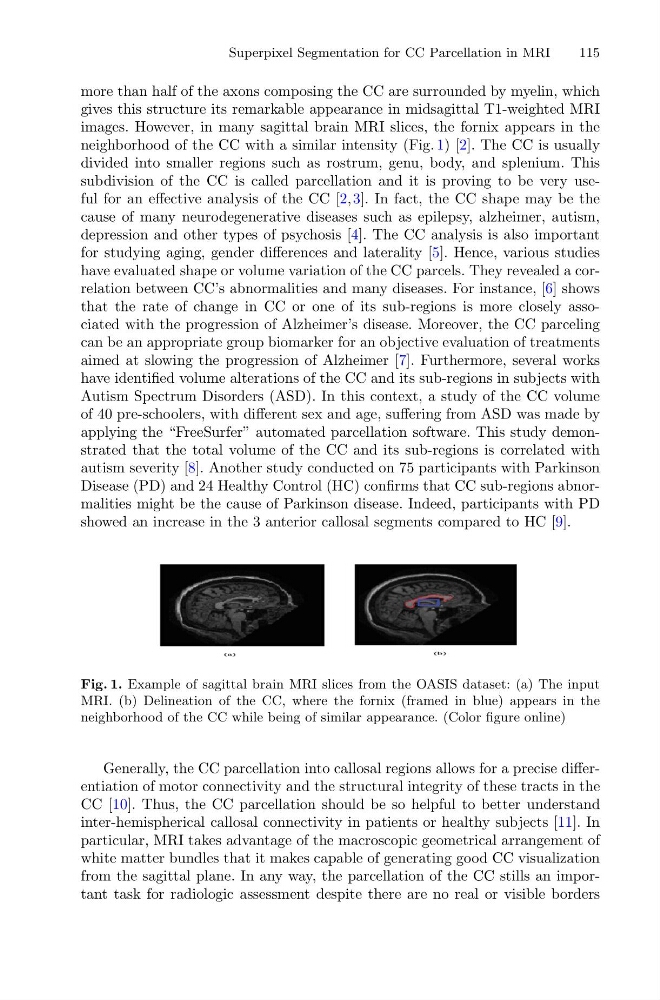
neighborhood of the CC with a similar intensity (Fig. 1) [2]. The CC is usually
divided into smaller regions such as rostrum, genu, body, and splenium. This
subdivision of the CC is called parcellation and it is proving to be very useful for an effective analysis of the CC [2,3]. In fact, the CC shape may be the
cause of many neurodegenerative diseases such as epilepsy, alzheimer, autism,
depression and other types of psychosis [4]. The CC analysis is also important
for studying aging, gender differences and laterality [5]. Hence, various studies
have evaluated shape or volume variation of the CC parcels. They revealed a correlation between CC’s abnormalities and many diseases. For instance, [6] shows
that the rate of change in CC or one of its sub-regions is more closely associated with the progression of Alzheimer’s disease. Moreover, the CC parceling
can be an appropriate group biomarker for an objective evaluation of treatments
aimed at slowing the progression of Alzheimer [7]. Furthermore, several works
have identified volume alterations of the CC and its sub-regions in subjects with
Autism Spectrum Disorders (ASD). In this context, a study of the CC volume
of 40 pre-schoolers, with different sex and age, suffering from ASD was made by
applying the “FreeSurfer” automated parcellation software. This study demonstrated that the total volume of the CC and its sub-regions is correlated with
autism severity [8]. Another study conducted on 75 participants with Parkinson
Disease (PD) and 24 Healthy Control (HC) confirms that CC sub-regions abnormalities might be the cause of Parkinson disease. Indeed, participants with PD
showed an increase in the 3 anterior callosal segments compared to HC [9].
Fig. 1. Example of sagittal brain MRI slices from the OASIS dataset: (a) The input
MRI. (b) Delineation of the CC, where the fornix (framed in blue) appears in the
neighborhood of the CC while being of similar appearance. (Color figure online)
Generally, the CC parcellation into callosal regions allows for a precise differentiation of motor connectivity and the structural integrity of these tracts in the
CC [10]. Thus, the CC parcellation should be so helpful to better understand
inter-hemispherical callosal connectivity in patients or healthy subjects [11]. In
particular, MRI takes advantage of the macroscopic geometrical arrangement of
white matter bundles that it makes capable of generating good CC visualization
from the sagittal plane. In any way, the parcellation of the CC stills an important task for radiologic assessment despite there are no real or visible borders
115
more than half of the axons composing the CC are surrounded by myelin, which
gives this structure its remarkable appearance in midsagittal T1-weighted MRI
images. However, in many sagittal brain MRI slices, the fornix appears in the
neighborhood of the CC with a similar intensity (Fig. 1) [2]. The CC is usually
divided into smaller regions such as rostrum, genu, body, and splenium. This
subdivision of the CC is called parcellation and it is proving to be very useful for an effective analysis of the CC [2,3]. In fact, the CC shape may be the
cause of many neurodegenerative diseases such as epilepsy, alzheimer, autism,
depression and other types of psychosis [4]. The CC analysis is also important
for studying aging, gender differences and laterality [5]. Hence, various studies
have evaluated shape or volume variation of the CC parcels. They revealed a correlation between CC’s abnormalities and many diseases. For instance, [6] shows
that the rate of change in CC or one of its sub-regions is more closely associated with the progression of Alzheimer’s disease. Moreover, the CC parceling
can be an appropriate group biomarker for an objective evaluation of treatments
aimed at slowing the progression of Alzheimer [7]. Furthermore, several works
have identified volume alterations of the CC and its sub-regions in subjects with
Autism Spectrum Disorders (ASD). In this context, a study of the CC volume
of 40 pre-schoolers, with different sex and age, suffering from ASD was made by
applying the “FreeSurfer” automated parcellation software. This study demonstrated that the total volume of the CC and its sub-regions is correlated with
autism severity [8]. Another study conducted on 75 participants with Parkinson
Disease (PD) and 24 Healthy Control (HC) confirms that CC sub-regions abnormalities might be the cause of Parkinson disease. Indeed, participants with PD
showed an increase in the 3 anterior callosal segments compared to HC [9].
Fig. 1. Example of sagittal brain MRI slices from the OASIS dataset: (a) The input
MRI. (b) Delineation of the CC, where the fornix (framed in blue) appears in the
neighborhood of the CC while being of similar appearance. (Color figure online)
Generally, the CC parcellation into callosal regions allows for a precise differentiation of motor connectivity and the structural integrity of these tracts in the
CC [10]. Thus, the CC parcellation should be so helpful to better understand
inter-hemispherical callosal connectivity in patients or healthy subjects [11]. In
particular, MRI takes advantage of the macroscopic geometrical arrangement of
white matter bundles that it makes capable of generating good CC visualization
from the sagittal plane. In any way, the parcellation of the CC stills an important task for radiologic assessment despite there are no real or visible borders