

T.P. Wróbel et al.
524
In vivo Studies of Atherosclerotic Plaques
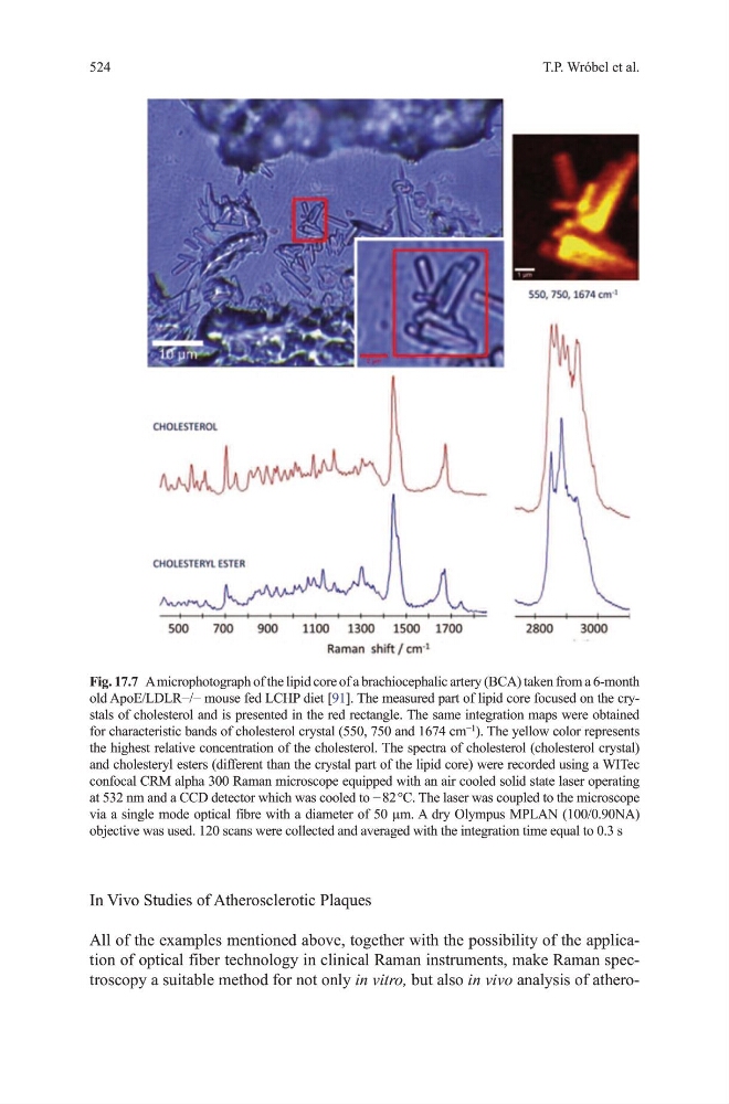
All of the examples mentioned above, together with the possibility of the application of optical fiber technology in clinical Raman instruments, make Raman spectroscopy a suitable method for not only in vitro, but also in vivo analysis of atheroFig. 17.7 A microphotograph of the lipid core of a brachiocephalic artery (BCA) taken from a 6-month
old ApoE/LDLR−/− mouse fed LCHP diet [91]. the measured part of lipid core focused on the crystals of cholesterol and is presented in the red rectangle. the same integration maps were obtained
for characteristic bands of cholesterol crystal (550, 750 and 1674 cm
−1
). the yellow color represents
the highest relative concentration of the cholesterol. the spectra of cholesterol (cholesterol crystal)
and cholesteryl esters (different than the crystal part of the lipid core) were recorded using a WItec
confocal CRm alpha 300 Raman microscope equipped with an air cooled solid state laser operating
at 532 nm and a CCD detector which was cooled to − 82 °C. The laser was coupled to the microscope
via a single mode optical fibre with a diameter of 50 µm. A dry olympus mPLAN (100/0.90NA)
objective was used. 120 scans were collected and averaged with the integration time equal to 0.3 s
524
In vivo Studies of Atherosclerotic Plaques
All of the examples mentioned above, together with the possibility of the application of optical fiber technology in clinical Raman instruments, make Raman spectroscopy a suitable method for not only in vitro, but also in vivo analysis of atheroFig. 17.7 A microphotograph of the lipid core of a brachiocephalic artery (BCA) taken from a 6-month
old ApoE/LDLR−/− mouse fed LCHP diet [91]. the measured part of lipid core focused on the crystals of cholesterol and is presented in the red rectangle. the same integration maps were obtained
for characteristic bands of cholesterol crystal (550, 750 and 1674 cm
−1
). the yellow color represents
the highest relative concentration of the cholesterol. the spectra of cholesterol (cholesterol crystal)
and cholesteryl esters (different than the crystal part of the lipid core) were recorded using a WItec
confocal CRm alpha 300 Raman microscope equipped with an air cooled solid state laser operating
at 532 nm and a CCD detector which was cooled to − 82 °C. The laser was coupled to the microscope
via a single mode optical fibre with a diameter of 50 µm. A dry olympus mPLAN (100/0.90NA)
objective was used. 120 scans were collected and averaged with the integration time equal to 0.3 s