

17 Vibrational Microspectroscopy for Analysis of Atherosclerotic Arteries
507
17.2.2 Atherosclerosis
Atherosclerosis (Atherothrombosis) is a major cause of ischaemic heart disease,
stroke and peripheral vascular disease and, therefore, a leading cause of death. Although it was believed that atherosclerotic plaque resulted from accumulation of
excessive plasma lipoproteins, progressively reducing the lumen of vessels it is
now clear that it is as a chronic inflammatory disease of the vascular wall that
is triggered and promoted by the development of endothelial dysfunction. these
mechanisms are far from being entirely understood. however, it is becoming increasingly clear that Low-density lipoproteins (LdL) and their subsequent enzymatic or non-enzymatic oxidation to oxidised LdL (ox-LdL) and accumulation
in subendothelial space triggers vascular inflammatory response the hallmark of
which is endothelial activation and dysfunction [27, 28]. the development of endothelial dysfunction results in infiltration of immune inflammatory cells (monocytes,
lymphocytes) to subendothelial space. monocytes differentiate into macrophages
(and/or dendritic cells) which phagocyte oxLdL and in consequence lose their mobility and converts to foam cells. Both foam cells and t-lymphocytes are detected in
fatty streaks— asymptomatic, easy reversible early predecessor of atherosclerotic
plaque. Formation of fatty streaks occurs in predilection sites with disturbed laminar
flow (i.e. arteries’ bifurcations) [29–34]. Atherosclerotic plaque formation involves
extensive production of extracellular matrix. Both foam cells, activated endothelium and vascular smooth muscle cells produce increased amount of extracellular
matrix in vascular wall which binds LdL subsequently beeing modified to oxLdL.
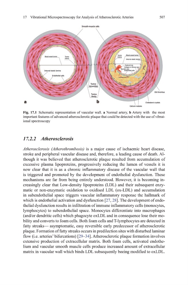
Fig. 17.1 Schematic representation of vascular wall. a Normal artery, b Artery with the most
important features of advanced atherosclerotic plaque that could be detected with the use of vibrational spectroscopy
507
17.2.2 Atherosclerosis
Atherosclerosis (Atherothrombosis) is a major cause of ischaemic heart disease,
stroke and peripheral vascular disease and, therefore, a leading cause of death. Although it was believed that atherosclerotic plaque resulted from accumulation of
excessive plasma lipoproteins, progressively reducing the lumen of vessels it is
now clear that it is as a chronic inflammatory disease of the vascular wall that
is triggered and promoted by the development of endothelial dysfunction. these
mechanisms are far from being entirely understood. however, it is becoming increasingly clear that Low-density lipoproteins (LdL) and their subsequent enzymatic or non-enzymatic oxidation to oxidised LdL (ox-LdL) and accumulation
in subendothelial space triggers vascular inflammatory response the hallmark of
which is endothelial activation and dysfunction [27, 28]. the development of endothelial dysfunction results in infiltration of immune inflammatory cells (monocytes,
lymphocytes) to subendothelial space. monocytes differentiate into macrophages
(and/or dendritic cells) which phagocyte oxLdL and in consequence lose their mobility and converts to foam cells. Both foam cells and t-lymphocytes are detected in
fatty streaks— asymptomatic, easy reversible early predecessor of atherosclerotic
plaque. Formation of fatty streaks occurs in predilection sites with disturbed laminar
flow (i.e. arteries’ bifurcations) [29–34]. Atherosclerotic plaque formation involves
extensive production of extracellular matrix. Both foam cells, activated endothelium and vascular smooth muscle cells produce increased amount of extracellular
matrix in vascular wall which binds LdL subsequently beeing modified to oxLdL.
Fig. 17.1 Schematic representation of vascular wall. a Normal artery, b Artery with the most
important features of advanced atherosclerotic plaque that could be detected with the use of vibrational spectroscopy