

489
16 Fourier Transform Infrared (Ft-Ir) Spectroscopic Imaging …
tween classes. typically, it is this combination of spectral features that are observed
in most biological samples and the gross distinction between different biological
or physiological conditions is significantly influenced by the differences in these
classes of compounds. the use of imaging enhances sensitivity to spectra differences by localizing signal. It is estimated that the sensitivity to differences likely
lies in the pg/pixel range for modern instruments.
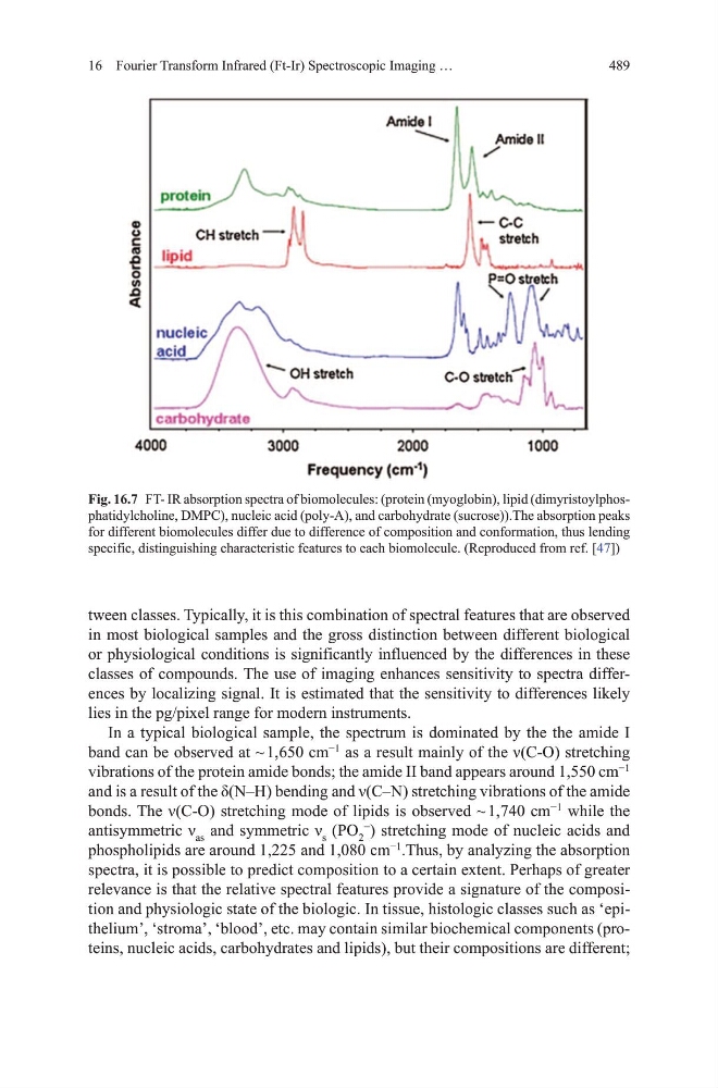
In a typical biological sample, the spectrum is dominated by the the amide I
band can be observed at ~ 1,650 cm
−1
as a result mainly of the ν(C-O) stretching
vibrations of the protein amide bonds; the amide II band appears around 1,550 cm
−1
and is a result of the δ(N–H) bending and ν(C–N) stretching vibrations of the amide
bonds. The ν(C-O) stretching mode of lipids is observed ~ 1,740 cm
−1
while the
antisymmetric ν as and symmetric ν s (Po 2
−
) stretching mode of nucleic acids and
phospholipids are around 1,225 and 1,080 cm
−1
.thus, by analyzing the absorption
spectra, it is possible to predict composition to a certain extent. Perhaps of greater
relevance is that the relative spectral features provide a signature of the composition and physiologic state of the biologic. In tissue, histologic classes such as ‘epithelium’, ‘stroma’, ‘blood’, etc. may contain similar biochemical components (proteins, nucleic acids, carbohydrates and lipids), but their compositions are different;
Fig. 16.7 Ft- IR absorption spectra of biomolecules: (protein (myoglobin), lipid (dimyristoylphosphatidylcholine, dmPC), nucleic acid (poly-A), and carbohydrate (sucrose)).the absorption peaks
for different biomolecules differ due to difference of composition and conformation, thus lending
specific, distinguishing characteristic features to each biomolecule. (Reproduced from ref. [47])
16 Fourier Transform Infrared (Ft-Ir) Spectroscopic Imaging …
tween classes. typically, it is this combination of spectral features that are observed
in most biological samples and the gross distinction between different biological
or physiological conditions is significantly influenced by the differences in these
classes of compounds. the use of imaging enhances sensitivity to spectra differences by localizing signal. It is estimated that the sensitivity to differences likely
lies in the pg/pixel range for modern instruments.
In a typical biological sample, the spectrum is dominated by the the amide I
band can be observed at ~ 1,650 cm
−1
as a result mainly of the ν(C-O) stretching
vibrations of the protein amide bonds; the amide II band appears around 1,550 cm
−1
and is a result of the δ(N–H) bending and ν(C–N) stretching vibrations of the amide
bonds. The ν(C-O) stretching mode of lipids is observed ~ 1,740 cm
−1
while the
antisymmetric ν as and symmetric ν s (Po 2
−
) stretching mode of nucleic acids and
phospholipids are around 1,225 and 1,080 cm
−1
.thus, by analyzing the absorption
spectra, it is possible to predict composition to a certain extent. Perhaps of greater
relevance is that the relative spectral features provide a signature of the composition and physiologic state of the biologic. In tissue, histologic classes such as ‘epithelium’, ‘stroma’, ‘blood’, etc. may contain similar biochemical components (proteins, nucleic acids, carbohydrates and lipids), but their compositions are different;
Fig. 16.7 Ft- IR absorption spectra of biomolecules: (protein (myoglobin), lipid (dimyristoylphosphatidylcholine, dmPC), nucleic acid (poly-A), and carbohydrate (sucrose)).the absorption peaks
for different biomolecules differ due to difference of composition and conformation, thus lending
specific, distinguishing characteristic features to each biomolecule. (Reproduced from ref. [47])