

449
15 FtIR Imaging of tissues: techniques and methods of Analysis
carried out by using fuzzy C-means clustering followed by uhCA. IR images were
collected from the five major tissue types: cervical stroma, squamous epithelium,
squamous cell carcinoma, dysplasia, and peritumoral inflammatory response. multivariate analysis in the region of 950–1480 cm
−1
discriminated all tissue layers with
some overlap. the cervical stroma and the area of inflammatory response along
with the transition layer between them contain a similar set of bands assigned to
proteins as the major constituent of these tissues. the variation in intensity of a band
at 1082 cm
−1
was found in spectra of all three tissue types and is a possible indicator
of inflammatory response. A higher intensity of the collagen band (a triplet centered
around 1232 cm
−1
) in the cervical stroma is found to be consistent with a lower
intensity of this band in the spectra of epithelium, while a cell layer between the
basal to parabasal tissues is characterized by a slightly increased absorbance near
1028 cm
−1
. despite the clear discrimination of the main components of the cervical tissue, the applied multivariate method did not categorize the basal cell layer,
dysplasia, and squamous cell carcinoma. only clustering in the spectral region of
1420–1480 cm
−1
showed some spectral discrimination of pathological states. this is
associated with shifting of a band assigned to the deformation vibration of the Ch 2
and Ch 3 groups from 1460 to 1452 cm
−1
, along with the decrease in its intensity,
which is maximum for the basal cell layer, medium for the dysplastic tissue, and
minimum for the squamous cell carcinoma [101].
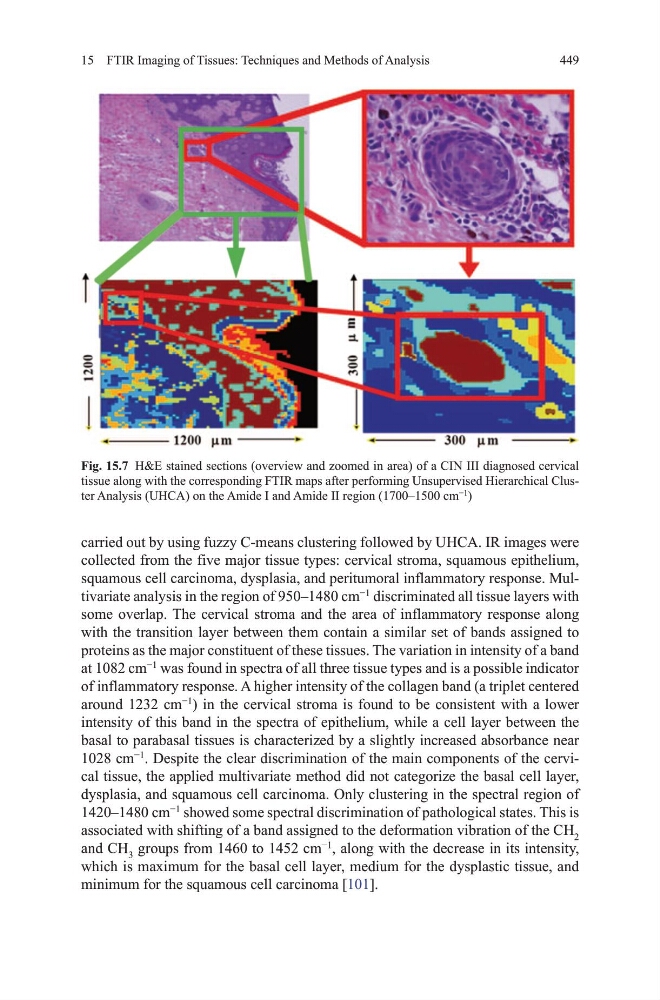
Fig. 15.7 h&E stained sections (overview and zoomed in area) of a CIN III diagnosed cervical
tissue along with the corresponding FtIR maps after performing unsupervised hierarchical Cluster Analysis (uhCA) on the Amide I and Amide II region (1700–1500 cm
−1
)
15 FtIR Imaging of tissues: techniques and methods of Analysis
carried out by using fuzzy C-means clustering followed by uhCA. IR images were
collected from the five major tissue types: cervical stroma, squamous epithelium,
squamous cell carcinoma, dysplasia, and peritumoral inflammatory response. multivariate analysis in the region of 950–1480 cm
−1
discriminated all tissue layers with
some overlap. the cervical stroma and the area of inflammatory response along
with the transition layer between them contain a similar set of bands assigned to
proteins as the major constituent of these tissues. the variation in intensity of a band
at 1082 cm
−1
was found in spectra of all three tissue types and is a possible indicator
of inflammatory response. A higher intensity of the collagen band (a triplet centered
around 1232 cm
−1
) in the cervical stroma is found to be consistent with a lower
intensity of this band in the spectra of epithelium, while a cell layer between the
basal to parabasal tissues is characterized by a slightly increased absorbance near
1028 cm
−1
. despite the clear discrimination of the main components of the cervical tissue, the applied multivariate method did not categorize the basal cell layer,
dysplasia, and squamous cell carcinoma. only clustering in the spectral region of
1420–1480 cm
−1
showed some spectral discrimination of pathological states. this is
associated with shifting of a band assigned to the deformation vibration of the Ch 2
and Ch 3 groups from 1460 to 1452 cm
−1
, along with the decrease in its intensity,
which is maximum for the basal cell layer, medium for the dysplastic tissue, and
minimum for the squamous cell carcinoma [101].
Fig. 15.7 h&E stained sections (overview and zoomed in area) of a CIN III diagnosed cervical
tissue along with the corresponding FtIR maps after performing unsupervised hierarchical Cluster Analysis (uhCA) on the Amide I and Amide II region (1700–1500 cm
−1
)