

433
15 FtIR Imaging of tissues: techniques and methods of Analysis
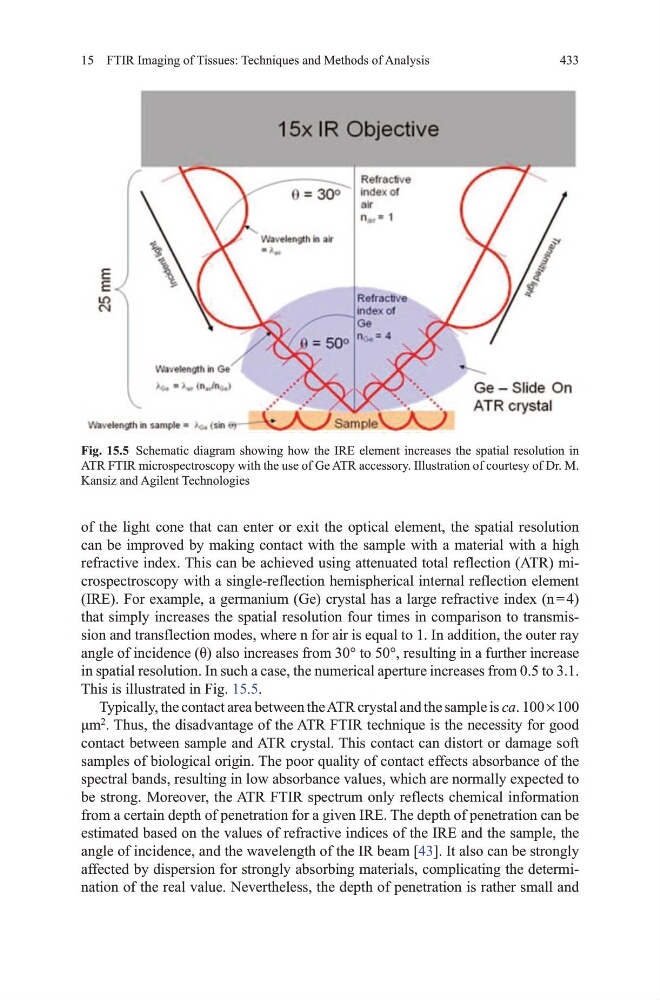
of the light cone that can enter or exit the optical element, the spatial resolution
can be improved by making contact with the sample with a material with a high
refractive index. this can be achieved using attenuated total reflection (AtR) microspectroscopy with a single-reflection hemispherical internal reflection element
(IRE). For example, a germanium (ge) crystal has a large refractive index (n = 4)
that simply increases the spatial resolution four times in comparison to transmission and transflection modes, where n for air is equal to 1. In addition, the outer ray
angle of incidence (θ) also increases from 30° to 50°, resulting in a further increase
in spatial resolution. In such a case, the numerical aperture increases from 0.5 to 3.1.
this is illustrated in Fig. 15.5.
typically, the contact area between the AtR crystal and the sample is ca. 100 × 100
μm
2
. thus, the disadvantage of the AtR FtIR technique is the necessity for good
contact between sample and AtR crystal. this contact can distort or damage soft
samples of biological origin. the poor quality of contact effects absorbance of the
spectral bands, resulting in low absorbance values, which are normally expected to
be strong. moreover, the AtR FtIR spectrum only reflects chemical information
from a certain depth of penetration for a given IRE. the depth of penetration can be
estimated based on the values of refractive indices of the IRE and the sample, the
angle of incidence, and the wavelength of the IR beam [43]. It also can be strongly
affected by dispersion for strongly absorbing materials, complicating the determination of the real value. Nevertheless, the depth of penetration is rather small and
Fig. 15.5 Schematic diagram showing how the IRE element increases the spatial resolution in
AtR FtIR microspectroscopy with the use of ge AtR accessory. Illustration of courtesy of dr. m.
Kansiz and Agilent technologies
15 FtIR Imaging of tissues: techniques and methods of Analysis
of the light cone that can enter or exit the optical element, the spatial resolution
can be improved by making contact with the sample with a material with a high
refractive index. this can be achieved using attenuated total reflection (AtR) microspectroscopy with a single-reflection hemispherical internal reflection element
(IRE). For example, a germanium (ge) crystal has a large refractive index (n = 4)
that simply increases the spatial resolution four times in comparison to transmission and transflection modes, where n for air is equal to 1. In addition, the outer ray
angle of incidence (θ) also increases from 30° to 50°, resulting in a further increase
in spatial resolution. In such a case, the numerical aperture increases from 0.5 to 3.1.
this is illustrated in Fig. 15.5.
typically, the contact area between the AtR crystal and the sample is ca. 100 × 100
μm
2
. thus, the disadvantage of the AtR FtIR technique is the necessity for good
contact between sample and AtR crystal. this contact can distort or damage soft
samples of biological origin. the poor quality of contact effects absorbance of the
spectral bands, resulting in low absorbance values, which are normally expected to
be strong. moreover, the AtR FtIR spectrum only reflects chemical information
from a certain depth of penetration for a given IRE. the depth of penetration can be
estimated based on the values of refractive indices of the IRE and the sample, the
angle of incidence, and the wavelength of the IR beam [43]. It also can be strongly
affected by dispersion for strongly absorbing materials, complicating the determination of the real value. Nevertheless, the depth of penetration is rather small and
Fig. 15.5 Schematic diagram showing how the IRE element increases the spatial resolution in
AtR FtIR microspectroscopy with the use of ge AtR accessory. Illustration of courtesy of dr. m.
Kansiz and Agilent technologies