

170
M. Pecul and W. Dzwolak
6.4 CD of Nucleic Acids
the local molecular chirality of nucleic acids originates from d-sugar molecules
(ribose in RNA and deoxyribose in dNA) which produce Cd bands in remote vacuum-uv. Although purine and pyrimidine bases have many electronic transitions
(mainly ππ* overlapping weaker nπ*) in an easily accessible UV range, their perfect planarity calls for additional conditions to be fulfilled before the corresponding
spectral bands could become Cd-active. Weak coupling to the sugar chiral center
allows only for a small Cd effect. however, the helical structure of dNA enables
building a strong and conformation-sensitive Cotton effect. the double-helix structure of dNA maintains large numbers of closely interacting bases arranged in a
chiral staircase. In an analogy to the case of induced Cd in near-uv Cd emerging
from chiral arrangement of achiral aromatic amino acid side chains in proteins, Cd
bands of helical dNA (or RNA) lying between 200 and 300 nm follow from asymmetry of the overall structure of the folded biopolymer. In case of far-uv protein
Cd, the spectral signal reflects primarily the conformation, and the amino acid sequence affects it only indirectly (given that partly overlapping but scarce aromatic
side chains cause only negligible contribution to uv absorption below 250 nm)
by promoting a particular secondary structure. Basically, the amide chromophore
is the same regardless of the sequence (one exception being proline). this is in
contrast to the more intricate case of dNA Cd where the contributing chromophore
(bases) and corresponding electronic transitions contributing to Cd change with
the sequence of the nucleic acid. hence fundamentally similar conformations of
dNA samples with different abundances of gC and At pairs will have distinct Cd
spectra. Because local interactions of transition dipoles between neighboring bases
affect chiroptical characteristics of folded nucleic acid, the sequence rather than just
nucleotide composition has to be taken into account when one attempts to simulate
the Cd spectrum of dNA with a known structure—a task remarkably more difficult
than computing protein far-uv Cd.
θ
λ
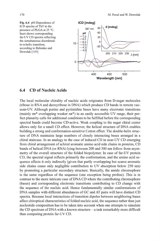
Fig. 6.4 ph-dependence of
ICd spectra of tht in the
presence of PLgA at 25 °C.
Inset shows corresponding
far-uv Cd spectra reflecting
the simultaneous disorderedto-α-helix transition,
according to Babenko and
dzwolak [109]
M. Pecul and W. Dzwolak
6.4 CD of Nucleic Acids
the local molecular chirality of nucleic acids originates from d-sugar molecules
(ribose in RNA and deoxyribose in dNA) which produce Cd bands in remote vacuum-uv. Although purine and pyrimidine bases have many electronic transitions
(mainly ππ* overlapping weaker nπ*) in an easily accessible UV range, their perfect planarity calls for additional conditions to be fulfilled before the corresponding
spectral bands could become Cd-active. Weak coupling to the sugar chiral center
allows only for a small Cd effect. however, the helical structure of dNA enables
building a strong and conformation-sensitive Cotton effect. the double-helix structure of dNA maintains large numbers of closely interacting bases arranged in a
chiral staircase. In an analogy to the case of induced Cd in near-uv Cd emerging
from chiral arrangement of achiral aromatic amino acid side chains in proteins, Cd
bands of helical dNA (or RNA) lying between 200 and 300 nm follow from asymmetry of the overall structure of the folded biopolymer. In case of far-uv protein
Cd, the spectral signal reflects primarily the conformation, and the amino acid sequence affects it only indirectly (given that partly overlapping but scarce aromatic
side chains cause only negligible contribution to uv absorption below 250 nm)
by promoting a particular secondary structure. Basically, the amide chromophore
is the same regardless of the sequence (one exception being proline). this is in
contrast to the more intricate case of dNA Cd where the contributing chromophore
(bases) and corresponding electronic transitions contributing to Cd change with
the sequence of the nucleic acid. hence fundamentally similar conformations of
dNA samples with different abundances of gC and At pairs will have distinct Cd
spectra. Because local interactions of transition dipoles between neighboring bases
affect chiroptical characteristics of folded nucleic acid, the sequence rather than just
nucleotide composition has to be taken into account when one attempts to simulate
the Cd spectrum of dNA with a known structure—a task remarkably more difficult
than computing protein far-uv Cd.
θ
λ
Fig. 6.4 ph-dependence of
ICd spectra of tht in the
presence of PLgA at 25 °C.
Inset shows corresponding
far-uv Cd spectra reflecting
the simultaneous disorderedto-α-helix transition,
according to Babenko and
dzwolak [109]