

168
M. Pecul and W. Dzwolak
of the native conformation if the native-disordered transitions were always taking
place cooperatively at every level of hierarchical architecture of protein native
state. however, detailed analysis of gradual denaturation of certain proteins, such
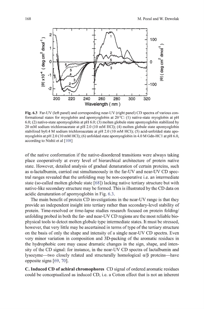
as α-lactalbumin, carried out simultaneously in the far-UV and near-UV CD spectral ranges revealed that the unfolding may be non-cooperative i.e. an intermediate
state (so-called molten globule state [68]) lacking native tertiary structure but with
native-like secondary structure may be formed. this is illustrated by the Cd data on
acidic denaturation of apomyoglobin in Fig. 6.3.
the main benefit of protein Cd investigations in the near-uv range is that they
provide an independent insight into tertiary rather than secondary-level stability of
protein. time-resolved or time-lapse studies research focused on protein folding/
unfolding probed in both the far- and near-uv Cd regions are the most reliable biophysical tools to detect molten globule type intermediate states. It must be stressed,
however, that very little may be ascertained in terms of type of the tertiary structure
on the basis of only the shape and intensity of a single near-uv Cd spectra. Even
very minor variation in composition and 3d-packing of the aromatic residues in
the hydrophobic core may cause dramatic changes in the sign, shape, and intensity of the Cd signal: for instance, in the near-uv Cd spectra of lactalbumin and
lysozyme—two closely related and structurally homological α/β proteins—have
opposite signs [69, 70].
C. Induced CD of achiral chromophores Cd signal of ordered aromatic residues
could be conceptualized as induced Cd, i.e. a Cotton effect that is not an inherent
Fig. 6.3 Far-uv (left panel) and corresponding near-uv (right panel) Cd spectra of various conformational states for myoglobin and apomyoglobin at 20 °C: (1) native-state myoglobin at ph
6.0; (2) native-state apomyoglobin at ph 6.0; (3) molten globule state apomyoglobin stabilized by
20 mm sodium trichloroacetate at ph 2.0 (10 mm hCl); (4) molten globule state apomyoglobin
stabilized by0.4 m sodium trichloroacetate at ph 2.0 (10 mm hCI); (5) acid-unfolded state apomyoglobin at ph 2.0 (10 mm hCl); (6) unfolded state apomyoglobin in 4.0 m gdn-hC1 at ph 6.0,
according to Nishii et al [108]
M. Pecul and W. Dzwolak
of the native conformation if the native-disordered transitions were always taking
place cooperatively at every level of hierarchical architecture of protein native
state. however, detailed analysis of gradual denaturation of certain proteins, such
as α-lactalbumin, carried out simultaneously in the far-UV and near-UV CD spectral ranges revealed that the unfolding may be non-cooperative i.e. an intermediate
state (so-called molten globule state [68]) lacking native tertiary structure but with
native-like secondary structure may be formed. this is illustrated by the Cd data on
acidic denaturation of apomyoglobin in Fig. 6.3.
the main benefit of protein Cd investigations in the near-uv range is that they
provide an independent insight into tertiary rather than secondary-level stability of
protein. time-resolved or time-lapse studies research focused on protein folding/
unfolding probed in both the far- and near-uv Cd regions are the most reliable biophysical tools to detect molten globule type intermediate states. It must be stressed,
however, that very little may be ascertained in terms of type of the tertiary structure
on the basis of only the shape and intensity of a single near-uv Cd spectra. Even
very minor variation in composition and 3d-packing of the aromatic residues in
the hydrophobic core may cause dramatic changes in the sign, shape, and intensity of the Cd signal: for instance, in the near-uv Cd spectra of lactalbumin and
lysozyme—two closely related and structurally homological α/β proteins—have
opposite signs [69, 70].
C. Induced CD of achiral chromophores Cd signal of ordered aromatic residues
could be conceptualized as induced Cd, i.e. a Cotton effect that is not an inherent
Fig. 6.3 Far-uv (left panel) and corresponding near-uv (right panel) Cd spectra of various conformational states for myoglobin and apomyoglobin at 20 °C: (1) native-state myoglobin at ph
6.0; (2) native-state apomyoglobin at ph 6.0; (3) molten globule state apomyoglobin stabilized by
20 mm sodium trichloroacetate at ph 2.0 (10 mm hCl); (4) molten globule state apomyoglobin
stabilized by0.4 m sodium trichloroacetate at ph 2.0 (10 mm hCI); (5) acid-unfolded state apomyoglobin at ph 2.0 (10 mm hCl); (6) unfolded state apomyoglobin in 4.0 m gdn-hC1 at ph 6.0,
according to Nishii et al [108]