

5 α-Amino Acids In Water: A Review of VCD and ROA Spectra
129
5.5.2.6 L-leucine
O
OH
NH 2
C
H 3
CH 3
Scheme 9 (S)-2-amino -4-methylpentanoic acid
VCD and ROA: Experiment and Calculations the vCd C*h stretching and
bending vibration bands of L-leucine in water were observed in early studies by the
Nafie group [147, 149]. Since then, no report on L-leucine vCd spectra was made
until the very recent study performed by the Xu group [215–217]. the IR spectra
of L-leucine in h 2 o and d 2 o were recorded under three representative ph conditions (1.0, 6.4, and 13); however, because of the poor solubility of L-leucine in
strongly acidic or basic solutions, the vCd spectra were taken only at near-neutral
ph/pd. As for L-serine and N-acetyl-L-cysteine in aqueous solutions [137, 213],
the conformational distribution of the dominant protonated, deprotonated, and
zwitterionic leucine species in water was studied in detail using quantum chemical calculations (B3LYP/6-311 + + g(d, p)). Additionally, the molecular dynamics
simulation of a leucine molecule solvated with 620 water molecules allowed for
the rational selection of zwitterionic-(water) n clusters for further dFt studies. the
authors found three protonated, three deprotonated, and three zwitterionic forms.
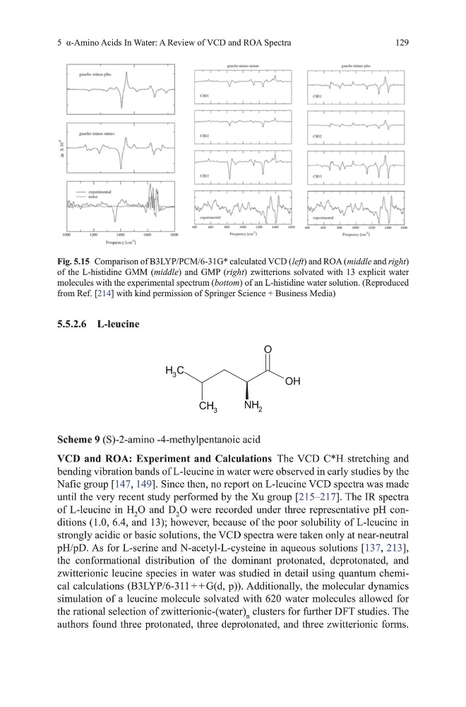
Fig. 5.15 Comparison of B3LYP/PCm/6-31g* calculated vCd ( left) and RoA ( middle and right)
of the L-histidine gmm (middle) and gmP (right) zwitterions solvated with 13 explicit water
molecules with the experimental spectrum (bottom) of an L-histidine water solution. (Reproduced
from Ref. [214] with kind permission of Springer Science + Business media)
129
5.5.2.6 L-leucine
O
OH
NH 2
C
H 3
CH 3
Scheme 9 (S)-2-amino -4-methylpentanoic acid
VCD and ROA: Experiment and Calculations the vCd C*h stretching and
bending vibration bands of L-leucine in water were observed in early studies by the
Nafie group [147, 149]. Since then, no report on L-leucine vCd spectra was made
until the very recent study performed by the Xu group [215–217]. the IR spectra
of L-leucine in h 2 o and d 2 o were recorded under three representative ph conditions (1.0, 6.4, and 13); however, because of the poor solubility of L-leucine in
strongly acidic or basic solutions, the vCd spectra were taken only at near-neutral
ph/pd. As for L-serine and N-acetyl-L-cysteine in aqueous solutions [137, 213],
the conformational distribution of the dominant protonated, deprotonated, and
zwitterionic leucine species in water was studied in detail using quantum chemical calculations (B3LYP/6-311 + + g(d, p)). Additionally, the molecular dynamics
simulation of a leucine molecule solvated with 620 water molecules allowed for
the rational selection of zwitterionic-(water) n clusters for further dFt studies. the
authors found three protonated, three deprotonated, and three zwitterionic forms.
Fig. 5.15 Comparison of B3LYP/PCm/6-31g* calculated vCd ( left) and RoA ( middle and right)
of the L-histidine gmm (middle) and gmP (right) zwitterions solvated with 13 explicit water
molecules with the experimental spectrum (bottom) of an L-histidine water solution. (Reproduced
from Ref. [214] with kind permission of Springer Science + Business media)