

J. C. Dobrowolsk et al.
108
5.5.2 Vibrational Chiroptical Studies on Particular Amino Acids
5.5.2.1 L-alanine
O
OH
C
H 3
NH 2
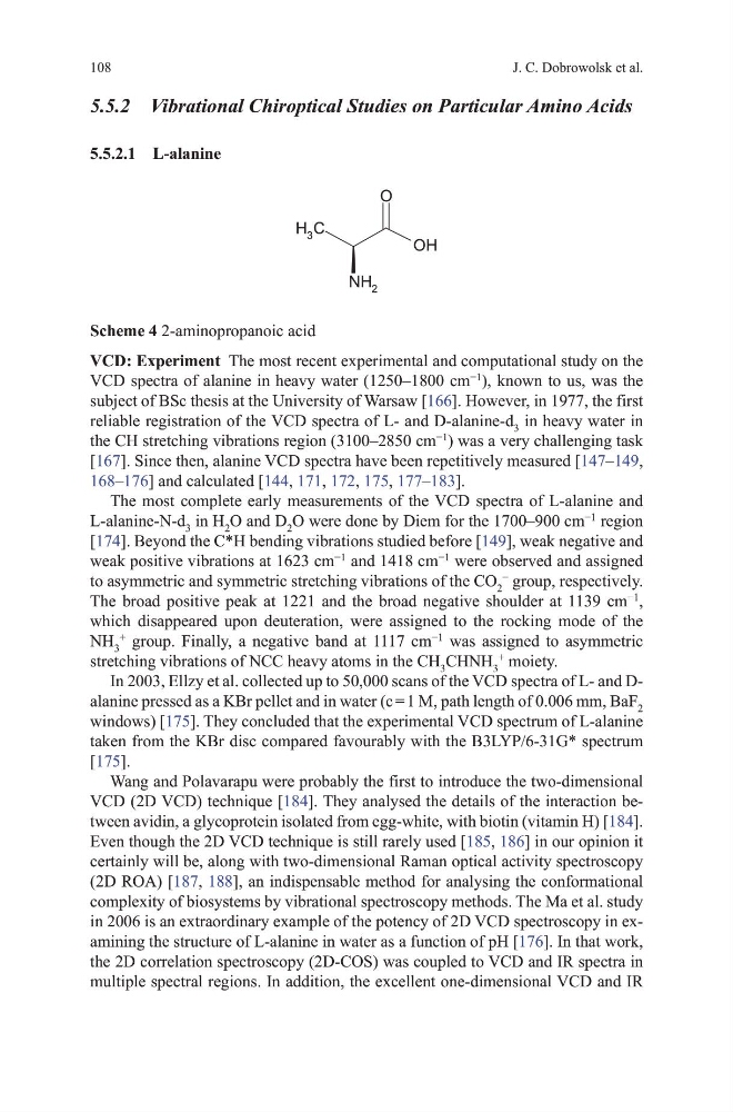
Scheme 4 2-aminopropanoic acid
VCD: Experiment the most recent experimental and computational study on the
vCd spectra of alanine in heavy water (1250–1800 cm
−1
), known to us, was the
subject of BSc thesis at the university of Warsaw [166]. however, in 1977, the first
reliable registration of the vCd spectra of L- and d-alanine-d 3 in heavy water in
the Ch stretching vibrations region (3100–2850 cm
−1
) was a very challenging task
[167]. Since then, alanine vCd spectra have been repetitively measured [147–149,
168–176] and calculated [144, 171, 172, 175, 177–183].
the most complete early measurements of the vCd spectra of L-alanine and
L-alanine-N-d 3 in h 2 o and d 2 o were done by diem for the 1700–900 cm
−1
region
[174]. Beyond the C*h bending vibrations studied before [149], weak negative and
weak positive vibrations at 1623 cm
−1
and 1418 cm
−1
were observed and assigned
to asymmetric and symmetric stretching vibrations of the Co 2
−
group, respectively.
the broad positive peak at 1221 and the broad negative shoulder at 1139 cm
−1
,
which disappeared upon deuteration, were assigned to the rocking mode of the
Nh 3
+
group. Finally, a negative band at 1117 cm
−1
was assigned to asymmetric
stretching vibrations of NCC heavy atoms in the Ch 3 ChNh 3
+
moiety.
In 2003, Ellzy et al. collected up to 50,000 scans of the vCd spectra of L- and dalanine pressed as a KBr pellet and in water (c = 1 m, path length of 0.006 mm, BaF 2
windows) [175]. they concluded that the experimental vCd spectrum of L-alanine
taken from the KBr disc compared favourably with the B3LYP/6-31g* spectrum
[175].
Wang and Polavarapu were probably the first to introduce the two-dimensional
vCd (2d vCd) technique [184]. they analysed the details of the interaction between avidin, a glycoprotein isolated from egg-white, with biotin (vitamin h) [184].
Even though the 2d vCd technique is still rarely used [185, 186] in our opinion it
certainly will be, along with two-dimensional Raman optical activity spectroscopy
(2d RoA) [187, 188], an indispensable method for analysing the conformational
complexity of biosystems by vibrational spectroscopy methods. the ma et al. study
in 2006 is an extraordinary example of the potency of 2d vCd spectroscopy in examining the structure of L-alanine in water as a function of ph [176]. In that work,
the 2d correlation spectroscopy (2d-CoS) was coupled to vCd and IR spectra in
multiple spectral regions. In addition, the excellent one-dimensional vCd and IR
108
5.5.2 Vibrational Chiroptical Studies on Particular Amino Acids
5.5.2.1 L-alanine
O
OH
C
H 3
NH 2
Scheme 4 2-aminopropanoic acid
VCD: Experiment the most recent experimental and computational study on the
vCd spectra of alanine in heavy water (1250–1800 cm
−1
), known to us, was the
subject of BSc thesis at the university of Warsaw [166]. however, in 1977, the first
reliable registration of the vCd spectra of L- and d-alanine-d 3 in heavy water in
the Ch stretching vibrations region (3100–2850 cm
−1
) was a very challenging task
[167]. Since then, alanine vCd spectra have been repetitively measured [147–149,
168–176] and calculated [144, 171, 172, 175, 177–183].
the most complete early measurements of the vCd spectra of L-alanine and
L-alanine-N-d 3 in h 2 o and d 2 o were done by diem for the 1700–900 cm
−1
region
[174]. Beyond the C*h bending vibrations studied before [149], weak negative and
weak positive vibrations at 1623 cm
−1
and 1418 cm
−1
were observed and assigned
to asymmetric and symmetric stretching vibrations of the Co 2
−
group, respectively.
the broad positive peak at 1221 and the broad negative shoulder at 1139 cm
−1
,
which disappeared upon deuteration, were assigned to the rocking mode of the
Nh 3
+
group. Finally, a negative band at 1117 cm
−1
was assigned to asymmetric
stretching vibrations of NCC heavy atoms in the Ch 3 ChNh 3
+
moiety.
In 2003, Ellzy et al. collected up to 50,000 scans of the vCd spectra of L- and dalanine pressed as a KBr pellet and in water (c = 1 m, path length of 0.006 mm, BaF 2
windows) [175]. they concluded that the experimental vCd spectrum of L-alanine
taken from the KBr disc compared favourably with the B3LYP/6-31g* spectrum
[175].
Wang and Polavarapu were probably the first to introduce the two-dimensional
vCd (2d vCd) technique [184]. they analysed the details of the interaction between avidin, a glycoprotein isolated from egg-white, with biotin (vitamin h) [184].
Even though the 2d vCd technique is still rarely used [185, 186] in our opinion it
certainly will be, along with two-dimensional Raman optical activity spectroscopy
(2d RoA) [187, 188], an indispensable method for analysing the conformational
complexity of biosystems by vibrational spectroscopy methods. the ma et al. study
in 2006 is an extraordinary example of the potency of 2d vCd spectroscopy in examining the structure of L-alanine in water as a function of ph [176]. In that work,
the 2d correlation spectroscopy (2d-CoS) was coupled to vCd and IR spectra in
multiple spectral regions. In addition, the excellent one-dimensional vCd and IR