

biological chromophores has become a common tool to gain new insights in their
intrinsic photophysical properties and the atomic-scale interactions that play a key
role for their functioning.
About 10 years ago, the first gas-phase absorption measurement of a
deprotonated GFP-model chromophore (a dimethyl derivative of pHBI, called
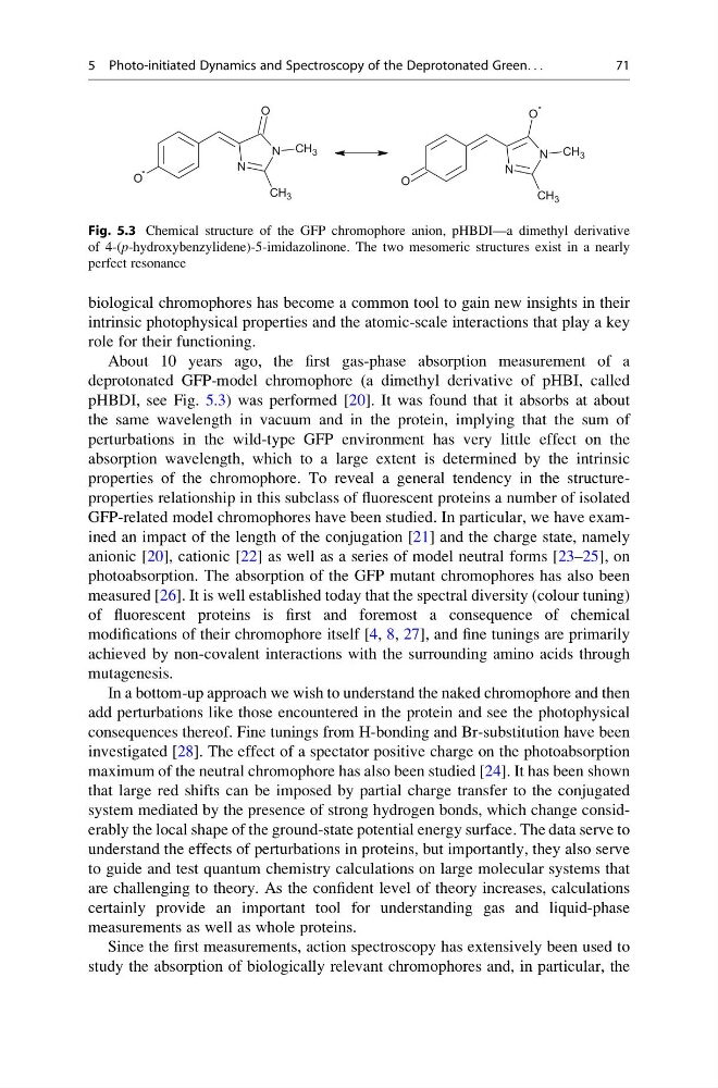
pHBDI, see Fig. 5.3) was performed [20]. It was found that it absorbs at about
the same wavelength in vacuum and in the protein, implying that the sum of
perturbations in the wild-type GFP environment has very little effect on the
absorption wavelength, which to a large extent is determined by the intrinsic
properties of the chromophore. To reveal a general tendency in the structureproperties relationship in this subclass of fluorescent proteins a number of isolated
GFP-related model chromophores have been studied. In particular, we have examined an impact of the length of the conjugation [21] and the charge state, namely
anionic [20], cationic [22] as well as a series of model neutral forms [23–25], on
photoabsorption. The absorption of the GFP mutant chromophores has also been
measured [26]. It is well established today that the spectral diversity (colour tuning)
of fluorescent proteins is first and foremost a consequence of chemical
modifications of their chromophore itself [4, 8, 27], and fine tunings are primarily
achieved by non-covalent interactions with the surrounding amino acids through
mutagenesis.
In a bottom-up approach we wish to understand the naked chromophore and then
add perturbations like those encountered in the protein and see the photophysical
consequences thereof. Fine tunings from H-bonding and Br-substitution have been
investigated [28]. The effect of a spectator positive charge on the photoabsorption
maximum of the neutral chromophore has also been studied [24]. It has been shown
that large red shifts can be imposed by partial charge transfer to the conjugated
system mediated by the presence of strong hydrogen bonds, which change considerably the local shape of the ground-state potential energy surface. The data serve to
understand the effects of perturbations in proteins, but importantly, they also serve
to guide and test quantum chemistry calculations on large molecular systems that
are challenging to theory. As the confident level of theory increases, calculations
certainly provide an important tool for understanding gas and liquid-phase
measurements as well as whole proteins.
Since the first measurements, action spectroscopy has extensively been used to
study the absorption of biologically relevant chromophores and, in particular, the
O
-
N
N
O
CH 3
CH 3
O
N
N
O
-
CH 3
CH 3
Fig. 5.3 Chemical structure of the GFP chromophore anion, pHBDI—a dimethyl derivative
of 4-(p-hydroxybenzylidene)-5-imidazolinone. The two mesomeric structures exist in a nearly
perfect resonance
5 Photo-initiated Dynamics and Spectroscopy of the Deprotonated Green. . .
71
intrinsic photophysical properties and the atomic-scale interactions that play a key
role for their functioning.
About 10 years ago, the first gas-phase absorption measurement of a
deprotonated GFP-model chromophore (a dimethyl derivative of pHBI, called
pHBDI, see Fig. 5.3) was performed [20]. It was found that it absorbs at about
the same wavelength in vacuum and in the protein, implying that the sum of
perturbations in the wild-type GFP environment has very little effect on the
absorption wavelength, which to a large extent is determined by the intrinsic
properties of the chromophore. To reveal a general tendency in the structureproperties relationship in this subclass of fluorescent proteins a number of isolated
GFP-related model chromophores have been studied. In particular, we have examined an impact of the length of the conjugation [21] and the charge state, namely
anionic [20], cationic [22] as well as a series of model neutral forms [23–25], on
photoabsorption. The absorption of the GFP mutant chromophores has also been
measured [26]. It is well established today that the spectral diversity (colour tuning)
of fluorescent proteins is first and foremost a consequence of chemical
modifications of their chromophore itself [4, 8, 27], and fine tunings are primarily
achieved by non-covalent interactions with the surrounding amino acids through
mutagenesis.
In a bottom-up approach we wish to understand the naked chromophore and then
add perturbations like those encountered in the protein and see the photophysical
consequences thereof. Fine tunings from H-bonding and Br-substitution have been
investigated [28]. The effect of a spectator positive charge on the photoabsorption
maximum of the neutral chromophore has also been studied [24]. It has been shown
that large red shifts can be imposed by partial charge transfer to the conjugated
system mediated by the presence of strong hydrogen bonds, which change considerably the local shape of the ground-state potential energy surface. The data serve to
understand the effects of perturbations in proteins, but importantly, they also serve
to guide and test quantum chemistry calculations on large molecular systems that
are challenging to theory. As the confident level of theory increases, calculations
certainly provide an important tool for understanding gas and liquid-phase
measurements as well as whole proteins.
Since the first measurements, action spectroscopy has extensively been used to
study the absorption of biologically relevant chromophores and, in particular, the
O
-
N
N
O
CH 3
CH 3
O
N
N
O
-
CH 3
CH 3
Fig. 5.3 Chemical structure of the GFP chromophore anion, pHBDI—a dimethyl derivative
of 4-(p-hydroxybenzylidene)-5-imidazolinone. The two mesomeric structures exist in a nearly
perfect resonance
5 Photo-initiated Dynamics and Spectroscopy of the Deprotonated Green. . .
71