

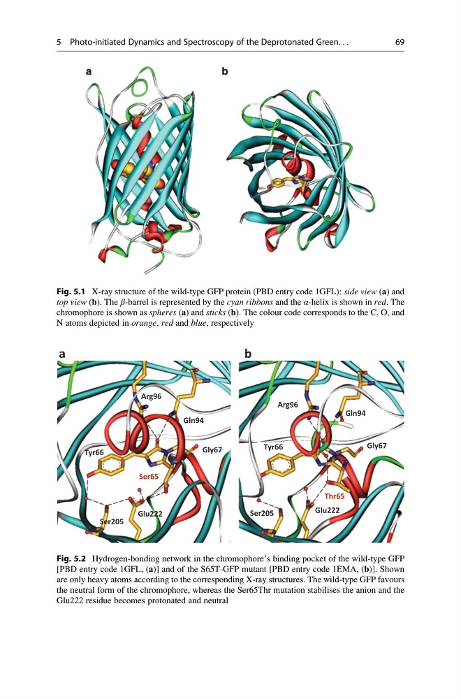
Fig. 5.1 X-ray structure of the wild-type GFP protein (PBD entry code 1GFL): side view (a) and
top view (b). The β-barrel is represented by the cyan ribbons and the α-helix is shown in red. The
chromophore is shown as spheres (a) and sticks (b). The colour code corresponds to the C, O, and
N atoms depicted in orange, red and blue, respectively
Tyr66
Gly67
Ser65
b
a
Glu222
Ser205
Arg96
Gln94
Tyr66
Gly67
Gln94
Arg96
Ser205
Glu222
Thr65
Fig. 5.2 Hydrogen-bonding network in the chromophore’s binding pocket of the wild-type GFP
[PBD entry code 1GFL, (a)] and of the S65T-GFP mutant [PBD entry code 1EMA, (b)]. Shown
are only heavy atoms according to the corresponding X-ray structures. The wild-type GFP favours
the neutral form of the chromophore, whereas the Ser65Thr mutation stabilises the anion and the
Glu222 residue becomes protonated and neutral
5 Photo-initiated Dynamics and Spectroscopy of the Deprotonated Green. . .
69
top view (b). The β-barrel is represented by the cyan ribbons and the α-helix is shown in red. The
chromophore is shown as spheres (a) and sticks (b). The colour code corresponds to the C, O, and
N atoms depicted in orange, red and blue, respectively
Tyr66
Gly67
Ser65
b
a
Glu222
Ser205
Arg96
Gln94
Tyr66
Gly67
Gln94
Arg96
Ser205
Glu222
Thr65
Fig. 5.2 Hydrogen-bonding network in the chromophore’s binding pocket of the wild-type GFP
[PBD entry code 1GFL, (a)] and of the S65T-GFP mutant [PBD entry code 1EMA, (b)]. Shown
are only heavy atoms according to the corresponding X-ray structures. The wild-type GFP favours
the neutral form of the chromophore, whereas the Ser65Thr mutation stabilises the anion and the
Glu222 residue becomes protonated and neutral
5 Photo-initiated Dynamics and Spectroscopy of the Deprotonated Green. . .
69