

blue or to the red. Only when we can account for the fate of each photoexcited
molecule, can we determine the actual absorption spectrum!
Another important issue is the effect of a single solvent molecule or counter ion
on the electronic structure of the biochromophore ion as mentioned above. The
location of the water could seriously affect the absorption or emission properties of
the chromophore/fluorophore. There is a sparse amount of data relating to this,
particularly experimental data. The motivation for looking at the effect of single
solvent molecules is of course that within hydrophobic protein pockets, there is
limited access to water, and often there are only one or two water molecules near to
the biochromophore. The advantage of experiments on ions is that such complexes
can be made from say ion-molecule reactions between the bare ions and gases or
even directly by electrospray ionisation, and they can then subsequently be studied
by mass spectroscopic means. Furthermore, water molecules can gradually be
added building up the complete solvation shell for comparison with absorption in
aqueous solution. Control, selectivity and “in a stepwise manner” are the key
words.
Most of the electronic spectroscopy experiments on biomolecular ions have been
done at room temperature, and another important direction would therefore be to
study cold ions that display much less spectral congestion and provide better
benchmarks for theory. Such experiments have successfully been done with regard
to vibrational spectroscopy employing 22-pole ion traps (pioneering work by Rizzo,
Boyarkin and their co-workers) [25, 26] based on the original trap design by Gerlich
[27]. The successful production of cold ions from the combination of electrospray
ionisation with cryo-cooled Paul traps by Wang, Johnson and their co-workers [28,
29] has also paved the way for studies of for example H 2 -tagged ions [29]; such
complexes are easily photodissociated with visible or ultraviolet light, thereby
circumpassing one of the inherent problems of action spectroscopy. Likewise,
Continetti and co-workers [30] have cryogenically cooled a linear electrostatic
ion beam trap for photoelectron-photofragment coincidence spectroscopy. Finally,
storage rings cooled down to a few Kelvin or at liquid nitrogen temperatures also
provide new interesting avenues along this direction. Such devices are built or
under construction in Stockholm, Heidelberg, and Tokyo [31–33]. Spectroscopy of
cold ions also provides direct information on the lifetimes of the excited states
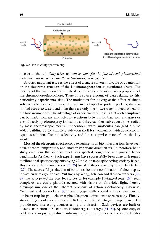
Fig. 2.7 Ion mobility spectrometry
16
S.B. Nielsen
molecule, can we determine the actual absorption spectrum!
Another important issue is the effect of a single solvent molecule or counter ion
on the electronic structure of the biochromophore ion as mentioned above. The
location of the water could seriously affect the absorption or emission properties of
the chromophore/fluorophore. There is a sparse amount of data relating to this,
particularly experimental data. The motivation for looking at the effect of single
solvent molecules is of course that within hydrophobic protein pockets, there is
limited access to water, and often there are only one or two water molecules near to
the biochromophore. The advantage of experiments on ions is that such complexes
can be made from say ion-molecule reactions between the bare ions and gases or
even directly by electrospray ionisation, and they can then subsequently be studied
by mass spectroscopic means. Furthermore, water molecules can gradually be
added building up the complete solvation shell for comparison with absorption in
aqueous solution. Control, selectivity and “in a stepwise manner” are the key
words.
Most of the electronic spectroscopy experiments on biomolecular ions have been
done at room temperature, and another important direction would therefore be to
study cold ions that display much less spectral congestion and provide better
benchmarks for theory. Such experiments have successfully been done with regard
to vibrational spectroscopy employing 22-pole ion traps (pioneering work by Rizzo,
Boyarkin and their co-workers) [25, 26] based on the original trap design by Gerlich
[27]. The successful production of cold ions from the combination of electrospray
ionisation with cryo-cooled Paul traps by Wang, Johnson and their co-workers [28,
29] has also paved the way for studies of for example H 2 -tagged ions [29]; such
complexes are easily photodissociated with visible or ultraviolet light, thereby
circumpassing one of the inherent problems of action spectroscopy. Likewise,
Continetti and co-workers [30] have cryogenically cooled a linear electrostatic
ion beam trap for photoelectron-photofragment coincidence spectroscopy. Finally,
storage rings cooled down to a few Kelvin or at liquid nitrogen temperatures also
provide new interesting avenues along this direction. Such devices are built or
under construction in Stockholm, Heidelberg, and Tokyo [31–33]. Spectroscopy of
cold ions also provides direct information on the lifetimes of the excited states
Fig. 2.7 Ion mobility spectrometry
16
S.B. Nielsen