

An additional fragment ion that is base-independent, but is not observed for all
parent ions, has m/z ¼ 151. This fragment was also found in CID experiments [34]
but has not yet been identified. Its independence of the identity of the nucleobase
indicates that it is a phosphate/sugar fragment. Finally, a fragment ion that was seen
only for the parent ion [AMP–H]
À , has m/z ¼ 139 [27, 28, 31]. The low abundance
of this ion, combined with the fact that it is not common to any other parent ion,
suggests that it may not be of much significance. No assignment has been offered
for this fragment ion so far.
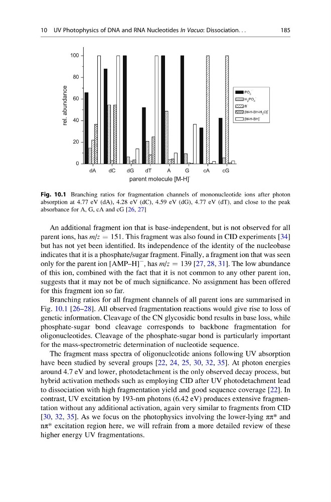
Branching ratios for all fragment channels of all parent ions are summarised in
Fig. 10.1 [26–28]. All observed fragmentation reactions would give rise to loss of
genetic information. Cleavage of the CN glycosidic bond results in base loss, while
phosphate-sugar bond cleavage corresponds to backbone fragmentation for
oligonucleotides. Cleavage of the phosphate-sugar bond is particularly important
for the mass-spectrometric determination of nucleotide sequence.
The fragment mass spectra of oligonucleotide anions following UV absorption
have been studied by several groups [22, 24, 25, 30, 32, 35]. At photon energies
around 4.7 eV and lower, photodetachment is the only observed decay process, but
hybrid activation methods such as employing CID after UV photodetachment lead
to dissociation with high fragmentation yield and good sequence coverage [22]. In
contrast, UV excitation by 193-nm photons (6.42 eV) produces extensive fragmentation without any additional activation, again very similar to fragments from CID
[30, 32, 35]. As we focus on the photophysics involving the lower-lying ππ* and
nπ* excitation region here, we will refrain from a more detailed review of these
higher energy UV fragmentations.
0
20
40
60
80
100
rel. abundance
parent molecule [M-H]
-
PO 3
-
H 2 PO 4
-
B
-
[M-H-BH-H 2 O]
-
[M-H-BH]
-
dA
dC
dG
dT
A
G
cA
cG
Fig. 10.1 Branching ratios for fragmentation channels of mononucleotide ions after photon
absorption at 4.77 eV (dA), 4.28 eV (dC), 4.59 eV (dG), 4.77 eV (dT), and close to the peak
absorbance for A, G, cA and cG [26, 27]
10 UV Photophysics of DNA and RNA Nucleotides In Vacuo: Dissociation. . .
185
parent ions, has m/z ¼ 151. This fragment was also found in CID experiments [34]
but has not yet been identified. Its independence of the identity of the nucleobase
indicates that it is a phosphate/sugar fragment. Finally, a fragment ion that was seen
only for the parent ion [AMP–H]
À , has m/z ¼ 139 [27, 28, 31]. The low abundance
of this ion, combined with the fact that it is not common to any other parent ion,
suggests that it may not be of much significance. No assignment has been offered
for this fragment ion so far.
Branching ratios for all fragment channels of all parent ions are summarised in
Fig. 10.1 [26–28]. All observed fragmentation reactions would give rise to loss of
genetic information. Cleavage of the CN glycosidic bond results in base loss, while
phosphate-sugar bond cleavage corresponds to backbone fragmentation for
oligonucleotides. Cleavage of the phosphate-sugar bond is particularly important
for the mass-spectrometric determination of nucleotide sequence.
The fragment mass spectra of oligonucleotide anions following UV absorption
have been studied by several groups [22, 24, 25, 30, 32, 35]. At photon energies
around 4.7 eV and lower, photodetachment is the only observed decay process, but
hybrid activation methods such as employing CID after UV photodetachment lead
to dissociation with high fragmentation yield and good sequence coverage [22]. In
contrast, UV excitation by 193-nm photons (6.42 eV) produces extensive fragmentation without any additional activation, again very similar to fragments from CID
[30, 32, 35]. As we focus on the photophysics involving the lower-lying ππ* and
nπ* excitation region here, we will refrain from a more detailed review of these
higher energy UV fragmentations.
0
20
40
60
80
100
rel. abundance
parent molecule [M-H]
-
PO 3
-
H 2 PO 4
-
B
-
[M-H-BH-H 2 O]
-
[M-H-BH]
-
dA
dC
dG
dT
A
G
cA
cG
Fig. 10.1 Branching ratios for fragmentation channels of mononucleotide ions after photon
absorption at 4.77 eV (dA), 4.28 eV (dC), 4.59 eV (dG), 4.77 eV (dT), and close to the peak
absorbance for A, G, cA and cG [26, 27]
10 UV Photophysics of DNA and RNA Nucleotides In Vacuo: Dissociation. . .
185