

five fragments ions, and if the intensity of four of these fragments is changing
when the probe laser is added while the intensity of the fifth fragment does not
change, one can deduce that this fifth fragment is not in competition with the
others and is a result of a totally different fragmentation mechanism or that a
specific conformer is producing this particular ion.
9.6.1 Tryptophan fs Dynamics
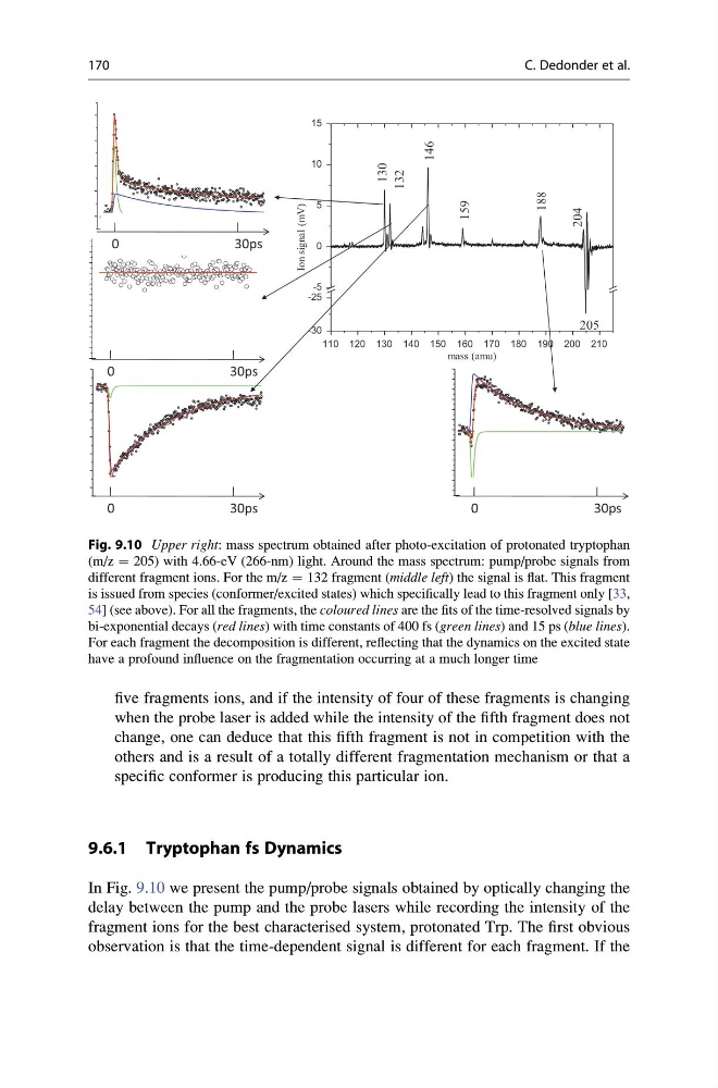
In Fig. 9.10 we present the pump/probe signals obtained by optically changing the
delay between the pump and the probe lasers while recording the intensity of the
fragment ions for the best characterised system, protonated Trp. The first obvious
observation is that the time-dependent signal is different for each fragment. If the
0
5000 10000 15000 20000 25000 30000 35000
delay 266/800nm (fs)
0
5000 10000 15000 20000 25000 30000 35000
delay 266/800nm (fs)
0
5000 10000 15000 20000 25000 30000 35000
delay 266/800nm (fs)
0
2000
4000
6000
8000
10000
fs
0
30ps
110 120 130 140 150 160 170 180 190 200 210
-30
-25
-5
0
5
10
15
205
130
132
146
159
204
188
Ion signal (mV)
mass (amu)
0
30ps
0
30ps
0
3 0 p s
Fig. 9.10 Upper right: mass spectrum obtained after photo-excitation of protonated tryptophan
(m/z ¼ 205) with 4.66-eV (266-nm) light. Around the mass spectrum: pump/probe signals from
different fragment ions. For the m/z ¼ 132 fragment (middle left) the signal is flat. This fragment
is issued from species (conformer/excited states) which specifically lead to this fragment only [33,
54] (see above). For all the fragments, the coloured lines are the fits of the time-resolved signals by
bi-exponential decays (red lines) with time constants of 400 fs (green lines) and 15 ps (blue lines).
For each fragment the decomposition is different, reflecting that the dynamics on the excited state
have a profound influence on the fragmentation occurring at a much longer time
170
C. Dedonder et al.
when the probe laser is added while the intensity of the fifth fragment does not
change, one can deduce that this fifth fragment is not in competition with the
others and is a result of a totally different fragmentation mechanism or that a
specific conformer is producing this particular ion.
9.6.1 Tryptophan fs Dynamics
In Fig. 9.10 we present the pump/probe signals obtained by optically changing the
delay between the pump and the probe lasers while recording the intensity of the
fragment ions for the best characterised system, protonated Trp. The first obvious
observation is that the time-dependent signal is different for each fragment. If the
0
5000 10000 15000 20000 25000 30000 35000
delay 266/800nm (fs)
0
5000 10000 15000 20000 25000 30000 35000
delay 266/800nm (fs)
0
5000 10000 15000 20000 25000 30000 35000
delay 266/800nm (fs)
0
2000
4000
6000
8000
10000
fs
0
30ps
110 120 130 140 150 160 170 180 190 200 210
-30
-25
-5
0
5
10
15
205
130
132
146
159
204
188
Ion signal (mV)
mass (amu)
0
30ps
0
30ps
0
3 0 p s
Fig. 9.10 Upper right: mass spectrum obtained after photo-excitation of protonated tryptophan
(m/z ¼ 205) with 4.66-eV (266-nm) light. Around the mass spectrum: pump/probe signals from
different fragment ions. For the m/z ¼ 132 fragment (middle left) the signal is flat. This fragment
is issued from species (conformer/excited states) which specifically lead to this fragment only [33,
54] (see above). For all the fragments, the coloured lines are the fits of the time-resolved signals by
bi-exponential decays (red lines) with time constants of 400 fs (green lines) and 15 ps (blue lines).
For each fragment the decomposition is different, reflecting that the dynamics on the excited state
have a profound influence on the fragmentation occurring at a much longer time
170
C. Dedonder et al.