

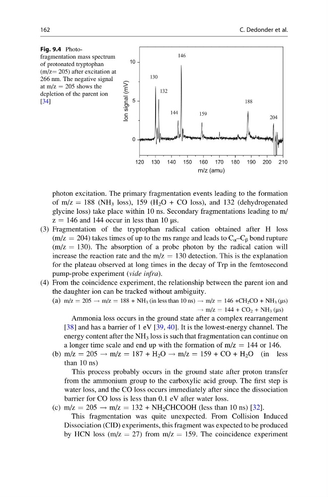
photon excitation. The primary fragmentation events leading to the formation
of m/z ¼ 188 (NH 3 loss), 159 (H 2 O + CO loss), and 132 (dehydrogenated
glycine loss) take place within 10 ns. Secondary fragmentations leading to m/
z ¼ 146 and 144 occur in less than 10 μs.
(3) Fragmentation of the tryptophan radical cation obtained after H loss
(m/z ¼ 204) takes times of up to the ms range and leads to C α –C β bond rupture
(m/z ¼ 130). The absorption of a probe photon by the radical cation will
increase the reaction rate and the m/z ¼ 130 detection. This is the explanation
for the plateau observed at long times in the decay of Trp in the femtosecond
pump-probe experiment (vide infra).
(4) From the coincidence experiment, the relationship between the parent ion and
the daughter ion can be tracked without ambiguity.
(a) m/z ¼ 205 ! m/z ¼ 188 + NH 3 (in less than 10 ns) ! m/z ¼ 146 +CH 2 CO + NH 3 (μs)
! m/z ¼ 144 + CO 2 + NH 3 (μs)
Ammonia loss occurs in the ground state after a complex rearrangement
[38] and has a barrier of 1 eV [39, 40]. It is the lowest-energy channel. The
energy content after the NH 3 loss is such that fragmentation can continue on
a longer time scale and end up with the formation of m/z ¼ 144 or 146.
(b) m/z ¼ 205 ! m/z ¼ 187 + H 2 O ! m/z ¼ 159 + CO + H 2 O (in less
than 10 ns)
This process probably occurs in the ground state after proton transfer
from the ammonium group to the carboxylic acid group. The first step is
water loss, and the CO loss occurs immediately after since the dissociation
barrier for CO loss is less than 0.1 eV after water loss.
(c) m/z ¼ 205 ! m/z ¼ 132 + NH 2 CHCOOH (less than 10 ns) [32].
This fragmentation was quite unexpected. From Collision Induced
Dissociation (CID) experiments, this fragment was expected to be produced
by HCN loss (m/z ¼ 27) from m/z ¼ 159. The coincidence experiment
120 130 140 150 160 170 180 190 200 210
0
5
10
144
130
132
146
159
204
188
Ion signal (mV)
m/z (amu)
Fig. 9.4 Photofragmentation mass spectrum
of protonated tryptophan
(m/z¼ 205) after excitation at
266 nm. The negative signal
at m/z ¼ 205 shows the
depletion of the parent ion
[34]
162
C. Dedonder et al.
of m/z ¼ 188 (NH 3 loss), 159 (H 2 O + CO loss), and 132 (dehydrogenated
glycine loss) take place within 10 ns. Secondary fragmentations leading to m/
z ¼ 146 and 144 occur in less than 10 μs.
(3) Fragmentation of the tryptophan radical cation obtained after H loss
(m/z ¼ 204) takes times of up to the ms range and leads to C α –C β bond rupture
(m/z ¼ 130). The absorption of a probe photon by the radical cation will
increase the reaction rate and the m/z ¼ 130 detection. This is the explanation
for the plateau observed at long times in the decay of Trp in the femtosecond
pump-probe experiment (vide infra).
(4) From the coincidence experiment, the relationship between the parent ion and
the daughter ion can be tracked without ambiguity.
(a) m/z ¼ 205 ! m/z ¼ 188 + NH 3 (in less than 10 ns) ! m/z ¼ 146 +CH 2 CO + NH 3 (μs)
! m/z ¼ 144 + CO 2 + NH 3 (μs)
Ammonia loss occurs in the ground state after a complex rearrangement
[38] and has a barrier of 1 eV [39, 40]. It is the lowest-energy channel. The
energy content after the NH 3 loss is such that fragmentation can continue on
a longer time scale and end up with the formation of m/z ¼ 144 or 146.
(b) m/z ¼ 205 ! m/z ¼ 187 + H 2 O ! m/z ¼ 159 + CO + H 2 O (in less
than 10 ns)
This process probably occurs in the ground state after proton transfer
from the ammonium group to the carboxylic acid group. The first step is
water loss, and the CO loss occurs immediately after since the dissociation
barrier for CO loss is less than 0.1 eV after water loss.
(c) m/z ¼ 205 ! m/z ¼ 132 + NH 2 CHCOOH (less than 10 ns) [32].
This fragmentation was quite unexpected. From Collision Induced
Dissociation (CID) experiments, this fragment was expected to be produced
by HCN loss (m/z ¼ 27) from m/z ¼ 159. The coincidence experiment
120 130 140 150 160 170 180 190 200 210
0
5
10
144
130
132
146
159
204
188
Ion signal (mV)
m/z (amu)
Fig. 9.4 Photofragmentation mass spectrum
of protonated tryptophan
(m/z¼ 205) after excitation at
266 nm. The negative signal
at m/z ¼ 205 shows the
depletion of the parent ion
[34]
162
C. Dedonder et al.