

[1–3]. The Trp excited state lifetime is also strongly dependent on the environment,
i.e., on the protein structure [1, 4–6].
The emission shift in proteins seems quite well understood [7] but the variation
of the excited state lifetime by more than two orders of magnitude is still not totally
elucidated [1, 8, 9]. In particular, it has been shown that Trp at low pH, in its
protonated form, has a very short lifetime [10]. To understand the basic properties
of the molecule, gas-phase studies along with ab-initio calculations are necessary
steps. The neutral aromatic amino acids have been characterised in the gas phase,
cooled in supersonic expansions where their spectroscopy is recorded using fluorescence [11, 12] or multi-photon ionisation detection schemes [5, 13–15]. These
molecules are very floppy species, and for example 12 conformers have been
detected for tyrosine, which all seem to have nanosecond excited state lifetimes.
It should be noted that conformers with very short lifetimes would not be detected
using nanosecond laser, and it has recently been shown in the case of neutral Phe
that the lifetime can change by one order of magnitude depending on the conformer
[16]. The variation of the lifetime is not due to variations of the oscillator strength
of the transition but to the appearance of non-radiative processes such as intersystem crossing, internal conversion or excited state photo-dissociation.
For the neutral systems, the spectroscopy of the excited states and the lifetimes
are well documented but the non-radiative processes are largely unknown. As an
example, it has been shown that phenol [17], the Tyr chromophore, loses an H atom
upon optical excitation but this reaction has not yet been investigated for tyrosine.
As will be seen in the following sections, a more complete story of the processes
following optical excitation can be tracked for protonated aromatic amino acids
(PAAAs), starting with spectroscopy with a tunable laser, to the excited states
dynamics, and followed by the primary and secondary fragmentations.
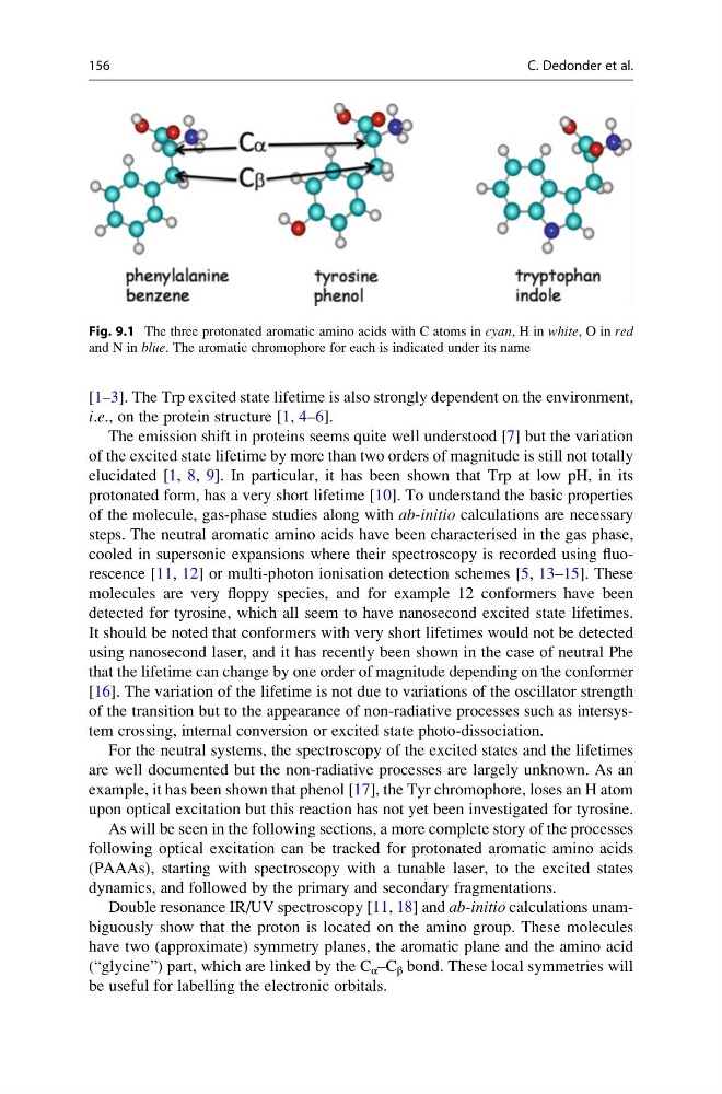
Double resonance IR/UV spectroscopy [11, 18] and ab-initio calculations unambiguously show that the proton is located on the amino group. These molecules
have two (approximate) symmetry planes, the aromatic plane and the amino acid
(“glycine”) part, which are linked by the C α –C β bond. These local symmetries will
be useful for labelling the electronic orbitals.
Fig. 9.1 The three protonated aromatic amino acids with C atoms in cyan, H in white, O in red
and N in blue. The aromatic chromophore for each is indicated under its name
156
C. Dedonder et al.
i.e., on the protein structure [1, 4–6].
The emission shift in proteins seems quite well understood [7] but the variation
of the excited state lifetime by more than two orders of magnitude is still not totally
elucidated [1, 8, 9]. In particular, it has been shown that Trp at low pH, in its
protonated form, has a very short lifetime [10]. To understand the basic properties
of the molecule, gas-phase studies along with ab-initio calculations are necessary
steps. The neutral aromatic amino acids have been characterised in the gas phase,
cooled in supersonic expansions where their spectroscopy is recorded using fluorescence [11, 12] or multi-photon ionisation detection schemes [5, 13–15]. These
molecules are very floppy species, and for example 12 conformers have been
detected for tyrosine, which all seem to have nanosecond excited state lifetimes.
It should be noted that conformers with very short lifetimes would not be detected
using nanosecond laser, and it has recently been shown in the case of neutral Phe
that the lifetime can change by one order of magnitude depending on the conformer
[16]. The variation of the lifetime is not due to variations of the oscillator strength
of the transition but to the appearance of non-radiative processes such as intersystem crossing, internal conversion or excited state photo-dissociation.
For the neutral systems, the spectroscopy of the excited states and the lifetimes
are well documented but the non-radiative processes are largely unknown. As an
example, it has been shown that phenol [17], the Tyr chromophore, loses an H atom
upon optical excitation but this reaction has not yet been investigated for tyrosine.
As will be seen in the following sections, a more complete story of the processes
following optical excitation can be tracked for protonated aromatic amino acids
(PAAAs), starting with spectroscopy with a tunable laser, to the excited states
dynamics, and followed by the primary and secondary fragmentations.
Double resonance IR/UV spectroscopy [11, 18] and ab-initio calculations unambiguously show that the proton is located on the amino group. These molecules
have two (approximate) symmetry planes, the aromatic plane and the amino acid
(“glycine”) part, which are linked by the C α –C β bond. These local symmetries will
be useful for labelling the electronic orbitals.
Fig. 9.1 The three protonated aromatic amino acids with C atoms in cyan, H in white, O in red
and N in blue. The aromatic chromophore for each is indicated under its name
156
C. Dedonder et al.