

part of the spectrum, which corresponds to a ~200 nm red-shift in comparison to the
first transition observed for neutral or deprotonated tryptophan-containing peptides
[1, 22]. This is due to a transition from an inner π orbital to the singly occupied π
HOMO [23].
Radical cations are conveniently formed in vacuo by electron transfer occurring
during collision-induced dissociation of ternary complexes composed of copper(II)
[24], a ligand and the peptide of interest. Capped AcGly 3 TrpNH 2 was used as a
model for small tryptophan-containing peptides. The optical spectrum of the radical
ion presented in Fig. 8.7b displays a broad absorption with maximum at 560 nm
[25]. This constitutes a redshift of about 300 nm as compared to molecular
tryptophan, and a redshift of about 100 nm as compared to the neutral tryptophan
radical (Fig. 8.7a). The band is due to a leading electronic transition of type π À2 π 0 .
Conclusion and Perspectives
One of the central results reported in this chapter is that among the multiple
possible de-excitation pathways after photoexcitation to electronic excited
states, relaxation of biomolecular polyanions is mainly achieved by electron
emission. Electron photodetachment is a fast process that occurs prior to relaxation into vibrational degrees of freedom. The electron photodetachment yield
can then be used to record gas-phase action spectra for systems as large as entire
proteins, without limitation of size due to energy redistribution into a large
number of modes.
Absorption spectra of peptides and proteins are sensitive to the electronic
configuration of the chromophores and to their environment. While UV–Vis
optical spectra directly depend on the electronic structure of chromophores, the
sensitivity of these spectra to protein conformation is more delicate to assess.
The results obtained on holo- and apo-myoglobin proteins are not conclusive. A
full interpretation of these spectra would require extensive theoretical efforts
that are beyond the scope of this chapter. A global strategy for the structural
characterisation of proteins and their complexes should involve different experimental approaches as well as theory. The action spectroscopy described here
could be complemented by ion mobility spectrometry (IMS) [26, 27], an
approach that we are now developing.
Another important aspect is the relationship between the dynamics of relaxation of the chromophore and its peptide or protein environment [28]. While
electron loss is the dominant process observed for polyanions, different
ions
fragment
tunable light visible
hv
light
UV
nm
hv
2
2H]
-
[M
-
e
2H]
-
[M
266
1
-
2
2H]
-
[M
+
·
-
+
·
-
+
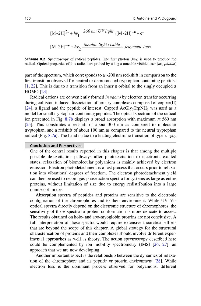
Scheme 8.2 Spectroscopy of radical peptides. The first photon (hν 1 ) is used to produce the
radical. Optical properties of this radical are probed by using a tuneable visible laser (hν 2 photon)
150
R. Antoine and P. Dugourd
first transition observed for neutral or deprotonated tryptophan-containing peptides
[1, 22]. This is due to a transition from an inner π orbital to the singly occupied π
HOMO [23].
Radical cations are conveniently formed in vacuo by electron transfer occurring
during collision-induced dissociation of ternary complexes composed of copper(II)
[24], a ligand and the peptide of interest. Capped AcGly 3 TrpNH 2 was used as a
model for small tryptophan-containing peptides. The optical spectrum of the radical
ion presented in Fig. 8.7b displays a broad absorption with maximum at 560 nm
[25]. This constitutes a redshift of about 300 nm as compared to molecular
tryptophan, and a redshift of about 100 nm as compared to the neutral tryptophan
radical (Fig. 8.7a). The band is due to a leading electronic transition of type π À2 π 0 .
Conclusion and Perspectives
One of the central results reported in this chapter is that among the multiple
possible de-excitation pathways after photoexcitation to electronic excited
states, relaxation of biomolecular polyanions is mainly achieved by electron
emission. Electron photodetachment is a fast process that occurs prior to relaxation into vibrational degrees of freedom. The electron photodetachment yield
can then be used to record gas-phase action spectra for systems as large as entire
proteins, without limitation of size due to energy redistribution into a large
number of modes.
Absorption spectra of peptides and proteins are sensitive to the electronic
configuration of the chromophores and to their environment. While UV–Vis
optical spectra directly depend on the electronic structure of chromophores, the
sensitivity of these spectra to protein conformation is more delicate to assess.
The results obtained on holo- and apo-myoglobin proteins are not conclusive. A
full interpretation of these spectra would require extensive theoretical efforts
that are beyond the scope of this chapter. A global strategy for the structural
characterisation of proteins and their complexes should involve different experimental approaches as well as theory. The action spectroscopy described here
could be complemented by ion mobility spectrometry (IMS) [26, 27], an
approach that we are now developing.
Another important aspect is the relationship between the dynamics of relaxation of the chromophore and its peptide or protein environment [28]. While
electron loss is the dominant process observed for polyanions, different
ions
fragment
tunable light visible
hv
light
UV
nm
hv
2
2H]
-
[M
-
e
2H]
-
[M
266
1
-
2
2H]
-
[M
+
·
-
+
·
-
+
Scheme 8.2 Spectroscopy of radical peptides. The first photon (hν 1 ) is used to produce the
radical. Optical properties of this radical are probed by using a tuneable visible laser (hν 2 photon)
150
R. Antoine and P. Dugourd