

described above, the vibrational energy dissipation pathway is of course not
accessible since there is no protein environment to interact with, and instead
vibrationally hot complexes are destined to dissociate. The assumption of complete
dissociation within the microsecond-time scale of the experiment seems to be a
good one.
7.3.1 Photoexcitation of Porphyrin Anions In Vacuo
The protoporphyrin IX anion (structure in Fig. 7.1c with one deprotonated carboxylic acid group) was also subjected to gas-phase spectroscopy experiments [44, 45].
In Fig. 7.16a the decay spectrum of PP anions after 430-nm photon excitation is
shown. The ions dissociated by loss of CO 2 , which only requires about half an eV.
Two exponential functions were needed to describe the decay of photoexcited ions,
with associated time constants of 67 μs and 0.87 ms. From power-dependence
measurements it was concluded that the whole decay was mainly due to the
0
2 0
4 0
6 0
8 0
10
1
10
2
10
3
10
4
Time after injection (ms)
Decay rate (arb. units)
a
b
10
1
10
2
10
3
10
4
Laser pulse fired
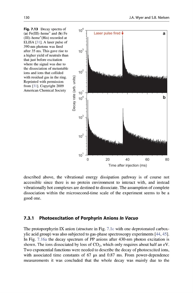
Fig. 7.13 Decay spectra of
(a) Fe(III)–heme
+ and (b) Fe
(III)–heme
+ (His) recorded at
ELISA [31]. A laser pulse of
390-nm photons was fired
after 35 ms. This gave rise to
a higher yield of neutrals than
that just before excitation
where the signal was due to
the dissociation of metastable
ions and ions that collided
with residual gas in the ring.
Reprinted with permission
from [31]. Copyright 2009
American Chemical Society
130
J.A. Wyer and S.B. Nielsen
accessible since there is no protein environment to interact with, and instead
vibrationally hot complexes are destined to dissociate. The assumption of complete
dissociation within the microsecond-time scale of the experiment seems to be a
good one.
7.3.1 Photoexcitation of Porphyrin Anions In Vacuo
The protoporphyrin IX anion (structure in Fig. 7.1c with one deprotonated carboxylic acid group) was also subjected to gas-phase spectroscopy experiments [44, 45].
In Fig. 7.16a the decay spectrum of PP anions after 430-nm photon excitation is
shown. The ions dissociated by loss of CO 2 , which only requires about half an eV.
Two exponential functions were needed to describe the decay of photoexcited ions,
with associated time constants of 67 μs and 0.87 ms. From power-dependence
measurements it was concluded that the whole decay was mainly due to the
0
2 0
4 0
6 0
8 0
10
1
10
2
10
3
10
4
Time after injection (ms)
Decay rate (arb. units)
a
b
10
1
10
2
10
3
10
4
Laser pulse fired
Fig. 7.13 Decay spectra of
(a) Fe(III)–heme
+ and (b) Fe
(III)–heme
+ (His) recorded at
ELISA [31]. A laser pulse of
390-nm photons was fired
after 35 ms. This gave rise to
a higher yield of neutrals than
that just before excitation
where the signal was due to
the dissociation of metastable
ions and ions that collided
with residual gas in the ring.
Reprinted with permission
from [31]. Copyright 2009
American Chemical Society
130
J.A. Wyer and S.B. Nielsen