

from mass spectrometry, which demonstrates the strength of FRET for following
structural changes of DNA.
The BODIPY-TMR dye was also used by Parks and co-workers [18–20] to
monitor conformational changes in the small unsolvated Trp-cage protein upon
heating. This protein only contains 20 amino-acid residues with a Trp residue
“caged” by three proline residues in a hydrophobic core. Here the Trp quenches
dye fluorescence when it is released from its cage. The quenching results from
intramolecular collisions between the excited dye and the Trp, resulting in electron
transfer from Trp to the half-filled orbital of the dye from where an electron was
photoexcited to a higher-lying orbital, followed by back transfer of the electron in
the excited-state orbital of the dye to Trp
+• [21]. This work showed that 3+ charge
state proteins more readily unfold than 2+ charge state proteins (see Fig. 6.2), partly
due to the greater Coulomb repulsion.
Conformational changes in dye-derivatised polyproline peptides, a β-hairpin
peptide, and vancomycin-peptide noncovalent complexes were studied in the
same way [20–22]. For polyproline peptides it was found that interactions between
the attached dye and charged residues led to variations in the fluorescence intensity
due to perturbation of the electronic levels of the dye (Stark effects). Furthermore,
as revealed from later work [23], the charge-transfer state between the dye and Trp,
D
À
T
+ , is lowered in energy in the vicinity of the electric field of a positively
charged arginine amino-acid residue (Fig. 6.3), explaining the fluorescence
quenching by charged residues. Hence the local environment of the dye-Trp pair
determines the fluorescence intensity. Clearly, a careful positioning of the dye, the
Trp, and charged residues allows one to associate a fluorescence change with a
specific structural change, and this technique therefore holds great promise in the
future for increasing our knowledge at a detailed level on the dynamics of protein
folding. Finally, Parks and co-workers [21] found from comparisons to solution-phase
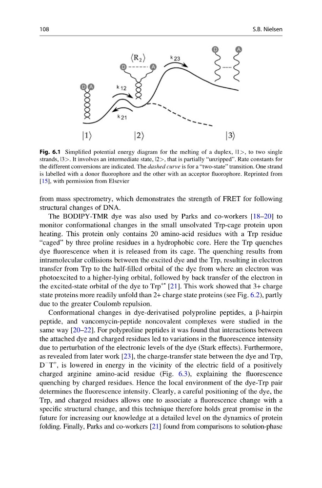
Fig. 6.1 Simplified potential energy diagram for the melting of a duplex, |1>, to two single
strands, |3>. It involves an intermediate state, |2>, that is partially “unzipped”. Rate constants for
the different conversions are indicated. The dashed curve is for a “two-state” transition. One strand
is labelled with a donor fluorophore and the other with an acceptor fluorophore. Reprinted from
[15], with permission from Elsevier
108
S.B. Nielsen
structural changes of DNA.
The BODIPY-TMR dye was also used by Parks and co-workers [18–20] to
monitor conformational changes in the small unsolvated Trp-cage protein upon
heating. This protein only contains 20 amino-acid residues with a Trp residue
“caged” by three proline residues in a hydrophobic core. Here the Trp quenches
dye fluorescence when it is released from its cage. The quenching results from
intramolecular collisions between the excited dye and the Trp, resulting in electron
transfer from Trp to the half-filled orbital of the dye from where an electron was
photoexcited to a higher-lying orbital, followed by back transfer of the electron in
the excited-state orbital of the dye to Trp
+• [21]. This work showed that 3+ charge
state proteins more readily unfold than 2+ charge state proteins (see Fig. 6.2), partly
due to the greater Coulomb repulsion.
Conformational changes in dye-derivatised polyproline peptides, a β-hairpin
peptide, and vancomycin-peptide noncovalent complexes were studied in the
same way [20–22]. For polyproline peptides it was found that interactions between
the attached dye and charged residues led to variations in the fluorescence intensity
due to perturbation of the electronic levels of the dye (Stark effects). Furthermore,
as revealed from later work [23], the charge-transfer state between the dye and Trp,
D
À
T
+ , is lowered in energy in the vicinity of the electric field of a positively
charged arginine amino-acid residue (Fig. 6.3), explaining the fluorescence
quenching by charged residues. Hence the local environment of the dye-Trp pair
determines the fluorescence intensity. Clearly, a careful positioning of the dye, the
Trp, and charged residues allows one to associate a fluorescence change with a
specific structural change, and this technique therefore holds great promise in the
future for increasing our knowledge at a detailed level on the dynamics of protein
folding. Finally, Parks and co-workers [21] found from comparisons to solution-phase
Fig. 6.1 Simplified potential energy diagram for the melting of a duplex, |1>, to two single
strands, |3>. It involves an intermediate state, |2>, that is partially “unzipped”. Rate constants for
the different conversions are indicated. The dashed curve is for a “two-state” transition. One strand
is labelled with a donor fluorophore and the other with an acceptor fluorophore. Reprinted from
[15], with permission from Elsevier
108
S.B. Nielsen