

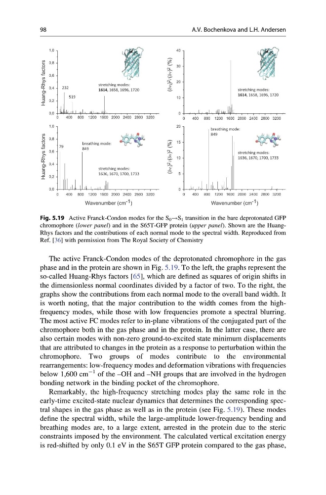
The active Franck-Condon modes of the deprotonated chromophore in the gas
phase and in the protein are shown in Fig. 5.19. To the left, the graphs represent the
so-called Huang-Rhys factors [65], which are defined as squares of origin shifts in
the dimensionless normal coordinates divided by a factor of two. To the right, the
graphs show the contributions from each normal mode to the overall band width. It
is worth noting, that the major contribution to the width comes from the highfrequency modes, while those with low frequencies promote a spectral blurring.
The most active FC modes refer to in-plane vibrations of the conjugated part of the
chromophore both in the gas phase and in the protein. In the latter case, there are
also certain modes with non-zero ground-to-excited state minimum displacements
that are attributed to changes in the protein as a response to perturbation within the
chromophore. Two groups of modes contribute to the environmental
rearrangements: low-frequency modes and deformation vibrations with frequencies
below 1,600 cm
À1 of the –OH and –NH groups that are involved in the hydrogen
bonding network in the binding pocket of the chromophore.
Remarkably, the high-frequency stretching modes play the same role in the
early-time excited-state nuclear dynamics that determines the corresponding spectral shapes in the gas phase as well as in the protein (see Fig. 5.19). These modes
define the spectral width, while the large-amplitude lower-frequency bending and
breathing modes are, to a large extent, arrested in the protein due to the steric
constraints imposed by the environment. The calculated vertical excitation energy
is red-shifted by only 0.1 eV in the S65T GFP protein compared to the gas phase,
0
400 800 1200 1600 2000 2400 2800 3200
0,0
0,2
0,4
0,6
0,8
1,0
Huang-Rhys factors
0
400 800 1200 1600 2000 2400 2800 3200
0
10
20
30
40
(
i ) 2
/
( ) 2
(%)
(
i ) 2
/
( ) 2
(%)
0
400 800 1200 1600 2000 2400 2800 3200
0,0
0,2
0,4
0,6
0,8
1,0
Huang-Rhys factors
Wavenumber (cm -1 )
0
400 800 1200 1600 2000 2400 2800 3200
0
5
10
15
20
Wavenumber (cm -1 )
stretching modes:
1636, 1670, 1700, 1733
stretching modes:
1636, 1670, 1700, 1733
breathing mode:
849
breathing mode:
849
stretching modes:
1614, 1658, 1696, 1720
79
232
519
stretching modes:
1614, 1658, 1696, 1720
Fig. 5.19 Active Franck-Condon modes for the S 0 !S 1 transition in the bare deprotonated GFP
chromophore (lower panel) and in the S65T-GFP protein (upper panel). Shown are the HuangRhys factors and the contributions of each normal mode to the spectral width. Reproduced from
Ref. [36] with permission from The Royal Society of Chemistry
98
A.V. Bochenkova and L.H. Andersen
phase and in the protein are shown in Fig. 5.19. To the left, the graphs represent the
so-called Huang-Rhys factors [65], which are defined as squares of origin shifts in
the dimensionless normal coordinates divided by a factor of two. To the right, the
graphs show the contributions from each normal mode to the overall band width. It
is worth noting, that the major contribution to the width comes from the highfrequency modes, while those with low frequencies promote a spectral blurring.
The most active FC modes refer to in-plane vibrations of the conjugated part of the
chromophore both in the gas phase and in the protein. In the latter case, there are
also certain modes with non-zero ground-to-excited state minimum displacements
that are attributed to changes in the protein as a response to perturbation within the
chromophore. Two groups of modes contribute to the environmental
rearrangements: low-frequency modes and deformation vibrations with frequencies
below 1,600 cm
À1 of the –OH and –NH groups that are involved in the hydrogen
bonding network in the binding pocket of the chromophore.
Remarkably, the high-frequency stretching modes play the same role in the
early-time excited-state nuclear dynamics that determines the corresponding spectral shapes in the gas phase as well as in the protein (see Fig. 5.19). These modes
define the spectral width, while the large-amplitude lower-frequency bending and
breathing modes are, to a large extent, arrested in the protein due to the steric
constraints imposed by the environment. The calculated vertical excitation energy
is red-shifted by only 0.1 eV in the S65T GFP protein compared to the gas phase,
0
400 800 1200 1600 2000 2400 2800 3200
0,0
0,2
0,4
0,6
0,8
1,0
Huang-Rhys factors
0
400 800 1200 1600 2000 2400 2800 3200
0
10
20
30
40
(
i ) 2
/
( ) 2
(%)
(
i ) 2
/
( ) 2
(%)
0
400 800 1200 1600 2000 2400 2800 3200
0,0
0,2
0,4
0,6
0,8
1,0
Huang-Rhys factors
Wavenumber (cm -1 )
0
400 800 1200 1600 2000 2400 2800 3200
0
5
10
15
20
Wavenumber (cm -1 )
stretching modes:
1636, 1670, 1700, 1733
stretching modes:
1636, 1670, 1700, 1733
breathing mode:
849
breathing mode:
849
stretching modes:
1614, 1658, 1696, 1720
79
232
519
stretching modes:
1614, 1658, 1696, 1720
Fig. 5.19 Active Franck-Condon modes for the S 0 !S 1 transition in the bare deprotonated GFP
chromophore (lower panel) and in the S65T-GFP protein (upper panel). Shown are the HuangRhys factors and the contributions of each normal mode to the spectral width. Reproduced from
Ref. [36] with permission from The Royal Society of Chemistry
98
A.V. Bochenkova and L.H. Andersen