

520
F. Gentile et al.
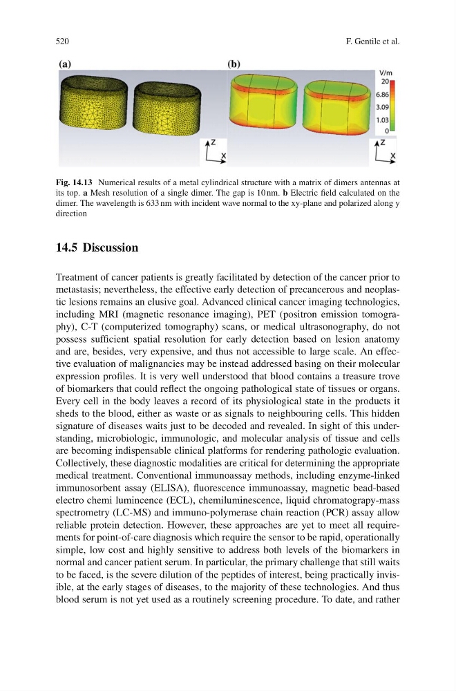
Fig. 14.13 Numerical results of a metal cylindrical structure with a matrix of dimers antennas at
its top. a Mesh resolution of a single dimer. The gap is 10 nm. b Electric field calculated on the
dimer. The wavelength is 633 nm with incident wave normal to the xy-plane and polarized along y
direction
14.5 Discussion
Treatment of cancer patients is greatly facilitated by detection of the cancer prior to
metastasis; nevertheless, the effective early detection of precancerous and neoplastic lesions remains an elusive goal. Advanced clinical cancer imaging technologies,
including MRI (magnetic resonance imaging), PET (positron emission tomography), C-T (computerized tomography) scans, or medical ultrasonography, do not
possess sufficient spatial resolution for early detection based on lesion anatomy
and are, besides, very expensive, and thus not accessible to large scale. An effective evaluation of malignancies may be instead addressed basing on their molecular
expression profiles. It is very well understood that blood contains a treasure trove
of biomarkers that could reflect the ongoing pathological state of tissues or organs.
Every cell in the body leaves a record of its physiological state in the products it
sheds to the blood, either as waste or as signals to neighbouring cells. This hidden
signature of diseases waits just to be decoded and revealed. In sight of this understanding, microbiologic, immunologic, and molecular analysis of tissue and cells
are becoming indispensable clinical platforms for rendering pathologic evaluation.
Collectively, these diagnostic modalities are critical for determining the appropriate
medical treatment. Conventional immunoassay methods, including enzyme-linked
immunosorbent assay (ELISA), fluorescence immunoassay, magnetic bead-based
electro chemi lumincence (ECL), chemiluminescence, liquid chromatograpy-mass
spectrometry (LC-MS) and immuno-polymerase chain reaction (PCR) assay allow
reliable protein detection. However, these approaches are yet to meet all requirements for point-of-care diagnosis which require the sensor to be rapid, operationally
simple, low cost and highly sensitive to address both levels of the biomarkers in
normal and cancer patient serum. In particular, the primary challenge that still waits
to be faced, is the severe dilution of the peptides of interest, being practically invisible, at the early stages of diseases, to the majority of these technologies. And thus
blood serum is not yet used as a routinely screening procedure. To date, and rather
F. Gentile et al.
Fig. 14.13 Numerical results of a metal cylindrical structure with a matrix of dimers antennas at
its top. a Mesh resolution of a single dimer. The gap is 10 nm. b Electric field calculated on the
dimer. The wavelength is 633 nm with incident wave normal to the xy-plane and polarized along y
direction
14.5 Discussion
Treatment of cancer patients is greatly facilitated by detection of the cancer prior to
metastasis; nevertheless, the effective early detection of precancerous and neoplastic lesions remains an elusive goal. Advanced clinical cancer imaging technologies,
including MRI (magnetic resonance imaging), PET (positron emission tomography), C-T (computerized tomography) scans, or medical ultrasonography, do not
possess sufficient spatial resolution for early detection based on lesion anatomy
and are, besides, very expensive, and thus not accessible to large scale. An effective evaluation of malignancies may be instead addressed basing on their molecular
expression profiles. It is very well understood that blood contains a treasure trove
of biomarkers that could reflect the ongoing pathological state of tissues or organs.
Every cell in the body leaves a record of its physiological state in the products it
sheds to the blood, either as waste or as signals to neighbouring cells. This hidden
signature of diseases waits just to be decoded and revealed. In sight of this understanding, microbiologic, immunologic, and molecular analysis of tissue and cells
are becoming indispensable clinical platforms for rendering pathologic evaluation.
Collectively, these diagnostic modalities are critical for determining the appropriate
medical treatment. Conventional immunoassay methods, including enzyme-linked
immunosorbent assay (ELISA), fluorescence immunoassay, magnetic bead-based
electro chemi lumincence (ECL), chemiluminescence, liquid chromatograpy-mass
spectrometry (LC-MS) and immuno-polymerase chain reaction (PCR) assay allow
reliable protein detection. However, these approaches are yet to meet all requirements for point-of-care diagnosis which require the sensor to be rapid, operationally
simple, low cost and highly sensitive to address both levels of the biomarkers in
normal and cancer patient serum. In particular, the primary challenge that still waits
to be faced, is the severe dilution of the peptides of interest, being practically invisible, at the early stages of diseases, to the majority of these technologies. And thus
blood serum is not yet used as a routinely screening procedure. To date, and rather