

5.12 Speckle-Optical Diagnostics of Muscle Activity and …
449
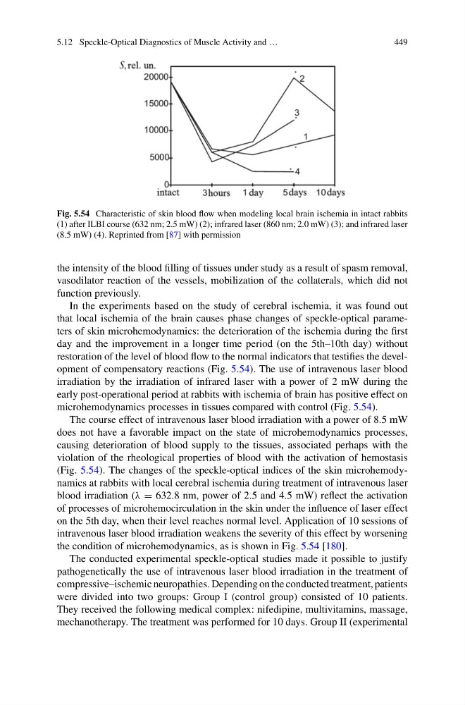
Fig. 5.54 Characteristic of skin blood flow when modeling local brain ischemia in intact rabbits
(1) after ILBI course (632 nm; 2.5 mW) (2); infrared laser (860 nm; 2.0 mW) (3); and infrared laser
(8.5 mW) (4). Reprinted from [87] with permission
the intensity of the blood filling of tissues under study as a result of spasm removal,
vasodilator reaction of the vessels, mobilization of the collaterals, which did not
function previously.
In the experiments based on the study of cerebral ischemia, it was found out
that local ischemia of the brain causes phase changes of speckle-optical parameters of skin microhemodynamics: the deterioration of the ischemia during the first
day and the improvement in a longer time period (on the 5th–10th day) without
restoration of the level of blood flow to the normal indicators that testifies the development of compensatory reactions (Fig. 5.54). The use of intravenous laser blood
irradiation by the irradiation of infrared laser with a power of 2 mW during the
early post-operational period at rabbits with ischemia of brain has positive effect on
microhemodynamics processes in tissues compared with control (Fig. 5.54).
The course effect of intravenous laser blood irradiation with a power of 8.5 mW
does not have a favorable impact on the state of microhemodynamics processes,
causing deterioration of blood supply to the tissues, associated perhaps with the
violation of the rheological properties of blood with the activation of hemostasis
(Fig. 5.54). The changes of the speckle-optical indices of the skin microhemodynamics at rabbits with local cerebral ischemia during treatment of intravenous laser
blood irradiation (λ = 632.8 nm, power of 2.5 and 4.5 mW) reflect the activation
of processes of microhemocirculation in the skin under the influence of laser effect
on the 5th day, when their level reaches normal level. Application of 10 sessions of
intravenous laser blood irradiation weakens the severity of this effect by worsening
the condition of microhemodynamics, as is shown in Fig. 5.54 [180].
The conducted experimental speckle-optical studies made it possible to justify
pathogenetically the use of intravenous laser blood irradiation in the treatment of
compressive–ischemic neuropathies. Depending on the conducted treatment, patients
were divided into two groups: Group I (control group) consisted of 10 patients.
They received the following medical complex: nifedipine, multivitamins, massage,
mechanotherapy. The treatment was performed for 10 days. Group II (experimental
449
Fig. 5.54 Characteristic of skin blood flow when modeling local brain ischemia in intact rabbits
(1) after ILBI course (632 nm; 2.5 mW) (2); infrared laser (860 nm; 2.0 mW) (3); and infrared laser
(8.5 mW) (4). Reprinted from [87] with permission
the intensity of the blood filling of tissues under study as a result of spasm removal,
vasodilator reaction of the vessels, mobilization of the collaterals, which did not
function previously.
In the experiments based on the study of cerebral ischemia, it was found out
that local ischemia of the brain causes phase changes of speckle-optical parameters of skin microhemodynamics: the deterioration of the ischemia during the first
day and the improvement in a longer time period (on the 5th–10th day) without
restoration of the level of blood flow to the normal indicators that testifies the development of compensatory reactions (Fig. 5.54). The use of intravenous laser blood
irradiation by the irradiation of infrared laser with a power of 2 mW during the
early post-operational period at rabbits with ischemia of brain has positive effect on
microhemodynamics processes in tissues compared with control (Fig. 5.54).
The course effect of intravenous laser blood irradiation with a power of 8.5 mW
does not have a favorable impact on the state of microhemodynamics processes,
causing deterioration of blood supply to the tissues, associated perhaps with the
violation of the rheological properties of blood with the activation of hemostasis
(Fig. 5.54). The changes of the speckle-optical indices of the skin microhemodynamics at rabbits with local cerebral ischemia during treatment of intravenous laser
blood irradiation (λ = 632.8 nm, power of 2.5 and 4.5 mW) reflect the activation
of processes of microhemocirculation in the skin under the influence of laser effect
on the 5th day, when their level reaches normal level. Application of 10 sessions of
intravenous laser blood irradiation weakens the severity of this effect by worsening
the condition of microhemodynamics, as is shown in Fig. 5.54 [180].
The conducted experimental speckle-optical studies made it possible to justify
pathogenetically the use of intravenous laser blood irradiation in the treatment of
compressive–ischemic neuropathies. Depending on the conducted treatment, patients
were divided into two groups: Group I (control group) consisted of 10 patients.
They received the following medical complex: nifedipine, multivitamins, massage,
mechanotherapy. The treatment was performed for 10 days. Group II (experimental