

266
3 Holographic Interferometry for Studying …
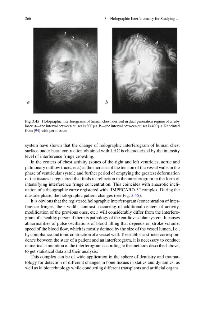
Fig. 3.45 Holographic interferograms of human chest, derived in dual generation regime of a ruby
laser: a—the interval between pulses is 300 μs; b—the interval between pulses is 400 μs. Reprinted
from [94] with permission
system have shown that the change of holographic interferogram of human chest
surface under heart contraction obtained with LHC is characterized by the intensity
level of interference fringe crowding.
In the centers of chest activity (zones of the right and left ventricles, aortic and
pulmonary outflow tracts, etc.) at the increase of the tension of the vessel walls in the
phase of ventricular systole and further period of emptying the greatest deformation
of the tissues is registered that finds its reflection in the interferogram in the form of
intensifying interference fringe concentration. This coincides with anacrotic inclination of a rheographic curve registered with “IMPECARD-3” complex. During the
diastole phase, the holographic pattern changes (see Fig. 3.45).
It is obvious that the registered holographic interferogram (concentration of interference fringes, their width, contrast, occurring of additional centers of activity,
modification of the previous ones, etc.) will considerably differ from the interferogram of a healthy person if there is pathology of the cardiovascular system. It causes
abnormalities of pulse oscillations of blood filling that depends on stroke volume,
speed of the blood flow, which is mostly defined by the size of the vessel lumen, i.e.,
by compliance and tonic contraction of a vessel wall. To establish a stricter correspondence between the state of a patient and an interferogram, it is necessary to conduct
numerical simulation of the interferogram according to the methods described above,
to get statistical data and their analysis.
This complex can be of wide application in the sphere of dentistry and traumatology for detection of different changes in bone tissues in statics and dynamics, as
well as in biotechnology while conducting different transplants and artificial organs.
3 Holographic Interferometry for Studying …
Fig. 3.45 Holographic interferograms of human chest, derived in dual generation regime of a ruby
laser: a—the interval between pulses is 300 μs; b—the interval between pulses is 400 μs. Reprinted
from [94] with permission
system have shown that the change of holographic interferogram of human chest
surface under heart contraction obtained with LHC is characterized by the intensity
level of interference fringe crowding.
In the centers of chest activity (zones of the right and left ventricles, aortic and
pulmonary outflow tracts, etc.) at the increase of the tension of the vessel walls in the
phase of ventricular systole and further period of emptying the greatest deformation
of the tissues is registered that finds its reflection in the interferogram in the form of
intensifying interference fringe concentration. This coincides with anacrotic inclination of a rheographic curve registered with “IMPECARD-3” complex. During the
diastole phase, the holographic pattern changes (see Fig. 3.45).
It is obvious that the registered holographic interferogram (concentration of interference fringes, their width, contrast, occurring of additional centers of activity,
modification of the previous ones, etc.) will considerably differ from the interferogram of a healthy person if there is pathology of the cardiovascular system. It causes
abnormalities of pulse oscillations of blood filling that depends on stroke volume,
speed of the blood flow, which is mostly defined by the size of the vessel lumen, i.e.,
by compliance and tonic contraction of a vessel wall. To establish a stricter correspondence between the state of a patient and an interferogram, it is necessary to conduct
numerical simulation of the interferogram according to the methods described above,
to get statistical data and their analysis.
This complex can be of wide application in the sphere of dentistry and traumatology for detection of different changes in bone tissues in statics and dynamics, as
well as in biotechnology while conducting different transplants and artificial organs.