

2.2 Holographic Study of Structural and Functional Characteristics …
111
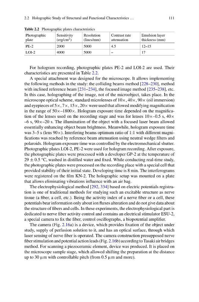
Table 2.2 Photographic plates characteristics
Photographic
plate
Sensitivity
(erg/cm 2 )
Resolution
(lines/mm)
Contrast rate
attenuation
Emulsion layer
thickness (mm)
PE-2
2000
5000
4.5
12–15
LOI-2
4000
5000
–
17
For hologram recording, photographic plates PE-2 and LOI-2 are used. Their
characteristics are presented in Table 2.2.
A special attachment was designed for the microscope. It allows implementing
the following methods in the study: the colliding beams method [228–230], method
with inclined reference beam [231–234], the focused image method [235–238], etc.
In this case, holographing of the image, not of the microobject, takes place. In the
microscope optical scheme, standard microlenses of 10×, 40×, 90× (oil immersion)
and eyepieces of 5×, 7×, 15×, 20× were used that allowed modifying magnification
in the range of 50×–1800×. Hologram exposure time depended on the magnification of the lenses used on the recording stage and was for lenses 10×–0.5 s, 40×
–6 s, 90×–20 s. The illumination of the object with a focused laser beam allowed
essentially enhancing object beam brightness. Meanwhile, hologram exposure time
was 3–5 s (lens 90×). Interfering beams optimum ratio of 1:1 with different magnifications was reached by reference beam attenuation using neutral wedge filters and
polaroids. Hologram exposure time was controlled by the electromechanical shutter.
Photographic plates LOI-2, PE-2 were used for hologram recording. After exposure,
the photographic plates were processed with a developer GP-2 at the temperature of
29 ± 0.5 °C, washed in distilled water and fixed. While conducting real-time study,
the photographic plates were processed on the recording place with a special cell that
provided stability of their initial state. Developing time is 8 min. The interferograms
were registered on the film KN-2. The holographic setup was mounted on a plate
that allows eliminating vibrations influence with an air bag.
The electrophysiological method [292, 334] based on electric potentials registration is one of traditional methods for studying such an excitable structure as nerve
tissue (a fiber, a cell, etc.). Being the activity index of a nerve fiber or a cell, these
potentials bear information only about ion fluxes alteration and do not give data about
the structure of fibers and cells. In these experiments, the electrophysiological part is
dedicated to nerve fiber activity control and contains an electrical stimulator ESU-2,
a special camera to fix the fiber, control oscillographs, a biopotential amplifier.
The camera (Fig. 2.16a) is a device, which provides fixation of the object under
study, supply of perfusion solution to it, and has an optical surface, through which
laser sensing of nerve fiber is operated. The camera construction presupposed nerve
fiber stimulation and potential action leads (Fig. 2.16b) according to Tasaki air bridges
method. For scanning a piezoceramic element, device was produced. It is placed on
the microscope sample stage, which allowed shifting the preparation at the distance
up to 30 μm with controllable pitch (from 0.5 μm and more).
111
Table 2.2 Photographic plates characteristics
Photographic
plate
Sensitivity
(erg/cm 2 )
Resolution
(lines/mm)
Contrast rate
attenuation
Emulsion layer
thickness (mm)
PE-2
2000
5000
4.5
12–15
LOI-2
4000
5000
–
17
For hologram recording, photographic plates PE-2 and LOI-2 are used. Their
characteristics are presented in Table 2.2.
A special attachment was designed for the microscope. It allows implementing
the following methods in the study: the colliding beams method [228–230], method
with inclined reference beam [231–234], the focused image method [235–238], etc.
In this case, holographing of the image, not of the microobject, takes place. In the
microscope optical scheme, standard microlenses of 10×, 40×, 90× (oil immersion)
and eyepieces of 5×, 7×, 15×, 20× were used that allowed modifying magnification
in the range of 50×–1800×. Hologram exposure time depended on the magnification of the lenses used on the recording stage and was for lenses 10×–0.5 s, 40×
–6 s, 90×–20 s. The illumination of the object with a focused laser beam allowed
essentially enhancing object beam brightness. Meanwhile, hologram exposure time
was 3–5 s (lens 90×). Interfering beams optimum ratio of 1:1 with different magnifications was reached by reference beam attenuation using neutral wedge filters and
polaroids. Hologram exposure time was controlled by the electromechanical shutter.
Photographic plates LOI-2, PE-2 were used for hologram recording. After exposure,
the photographic plates were processed with a developer GP-2 at the temperature of
29 ± 0.5 °C, washed in distilled water and fixed. While conducting real-time study,
the photographic plates were processed on the recording place with a special cell that
provided stability of their initial state. Developing time is 8 min. The interferograms
were registered on the film KN-2. The holographic setup was mounted on a plate
that allows eliminating vibrations influence with an air bag.
The electrophysiological method [292, 334] based on electric potentials registration is one of traditional methods for studying such an excitable structure as nerve
tissue (a fiber, a cell, etc.). Being the activity index of a nerve fiber or a cell, these
potentials bear information only about ion fluxes alteration and do not give data about
the structure of fibers and cells. In these experiments, the electrophysiological part is
dedicated to nerve fiber activity control and contains an electrical stimulator ESU-2,
a special camera to fix the fiber, control oscillographs, a biopotential amplifier.
The camera (Fig. 2.16a) is a device, which provides fixation of the object under
study, supply of perfusion solution to it, and has an optical surface, through which
laser sensing of nerve fiber is operated. The camera construction presupposed nerve
fiber stimulation and potential action leads (Fig. 2.16b) according to Tasaki air bridges
method. For scanning a piezoceramic element, device was produced. It is placed on
the microscope sample stage, which allowed shifting the preparation at the distance
up to 30 μm with controllable pitch (from 0.5 μm and more).